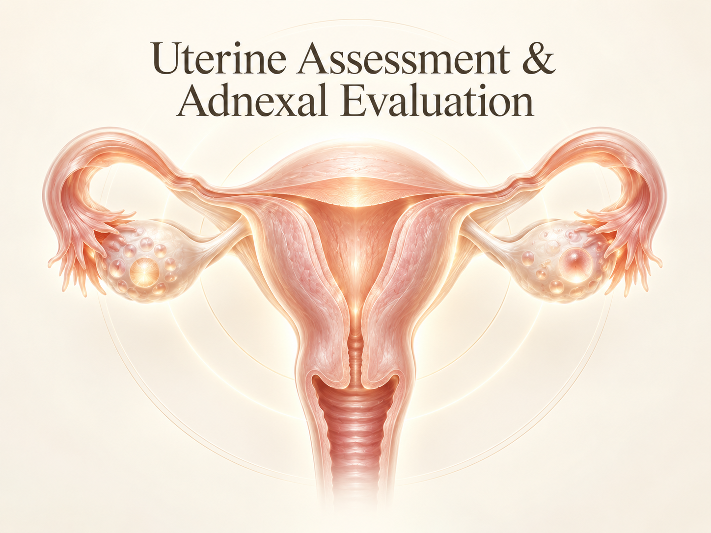
Uterine Assessment & Adnexal Evaluation
Evaluation of uterine size, shape, myometrium (fibroids/adenomyosis), endometrium, and systematic adnexal scanning.
This module is open to all medical professionals at no cost. Sign in with Google to unlock the interactive simulator.
100% Free & Open Access
Detailed Course Syllabus
Standard Transvaginal Gynecologic Survey
Transducer orientation, anteverted vs retroverted uterus, and scanning technique.
Myometrial Pathology: Fibroids vs Adenomyosis
FIGO fibroid classification (0–8), acoustic shadowing patterns, and junctional zone infiltration.
Endometrial Assessment & Sonohysterography
Triple-line pattern, postmenopausal bleeding cut-offs (< 4 mm), and saline infusion sonography.
IOTA Simple Rules & Ovarian Staging
Benign B-features, malignant M-features, and color Doppler scoring.
Learning Objectives
- Apply the MUSA (Morphological Uterus Sonographic Assessment) criteria for adenomyosis and leiomyomas.
- Delineate endometrial stripe abnormalities: polyps, hyperplasia, and endometrial carcinoma.
- Implement the IOTA (International Ovarian Tumor Analysis) Simple Rules for adnexal mass characterization.
- Differentiate benign functional cysts from mature teratomas, endometriomas, and cystadenomas.
Clinical Relevance
Standardized nomenclature in gynecological sonography reduces misinterpretation, prevents unnecessary laparotomies for benign cysts, and accelerates gynecologic oncologic triage.
Clinical Guidelines Aligned
- IOTA Consensus Guidelines on Ovarian Mass Characterization
- MUSA Consensus on Reporting Myometrial Pathology
Explore Other Courses
Sonoembryology Course (Weeks 1 to 11)
Interactive exploration of early pregnancy ultrasound findings, developmental landmarks, and pitfalls.
Detailed First-Trimester Scan (12w 0d to 13w 6d)
Early anatomical survey, nuchal translucency caliper calibration, nasal bone assessment, and aneuploidy risk calculation.
Second and Third Trimesters (≥16+0 weeks)
Systematic screening of fetal structural anatomy, biometry measurements, and placental assessment.