Sonoembryology Course (Weeks 1 to 11)
Interactive exploration of early pregnancy ultrasound findings, developmental landmarks, and pitfalls.
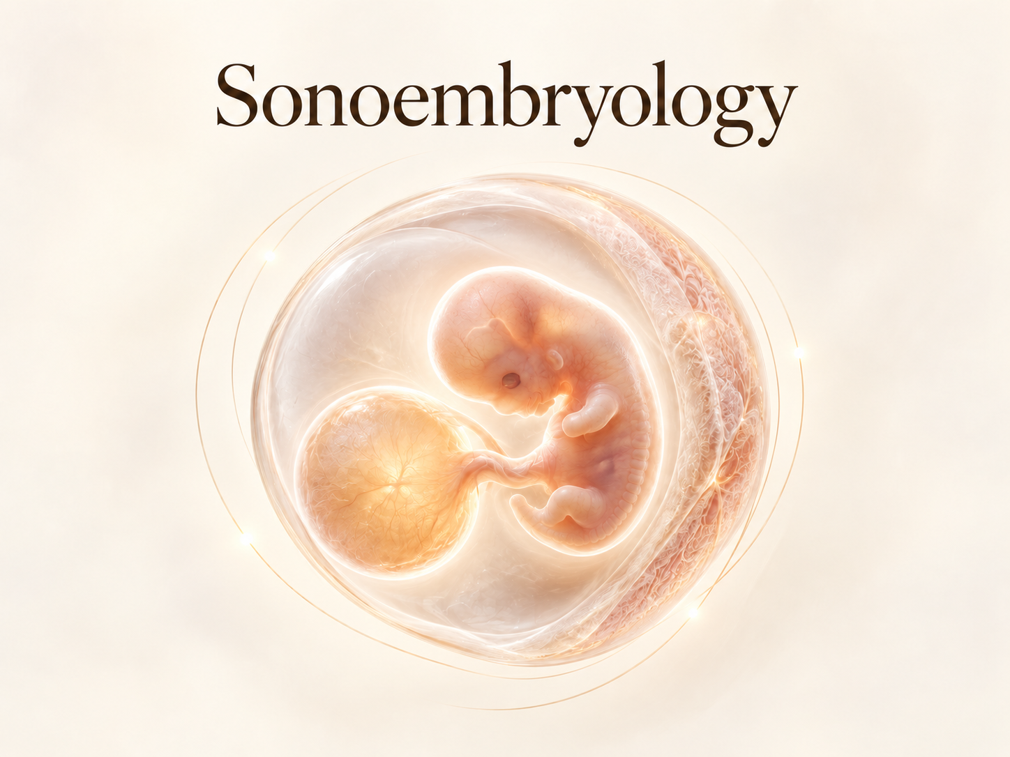
This module is open to all medical professionals at no cost. Sign in with Google to unlock the interactive simulator.
100% Free & Open Access
Sonoembryology Virtual Ultrasound Simulator
Early Embryonic & Cardiac Morphogenesis
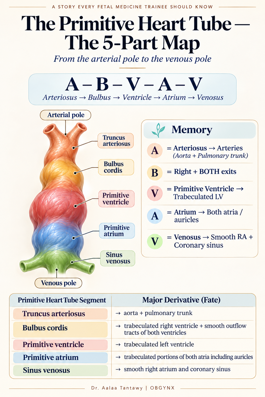
A – B – V – A – V
Arteriosus → Bulbus → Ventricle → Atrium → Venosus. The primitive tube undergoes normal rightward (dextral, D-) looping, rearranging these segments into the spatial relationships of the developing heart.
Truncus Arteriosus
The future great arterial outlet
The most cranial arterial part of the primitive heart tube. Contributes to the proximal great arterial outflow: Aorta + Pulmonary trunk.
A = Arteriosus → Arteries (Aorta + Pulmonary)
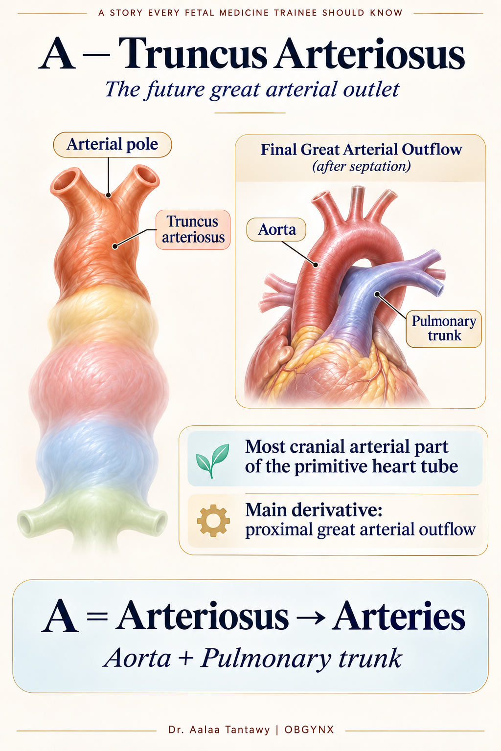 A — Truncus Arteriosus: Aorta & Pulmonary Trunk
Then the Heart Tube Loops (D-Looping = Rightward Looping)
This transforms a straight tube into a three-dimensional heart. The 3 key directional moves:
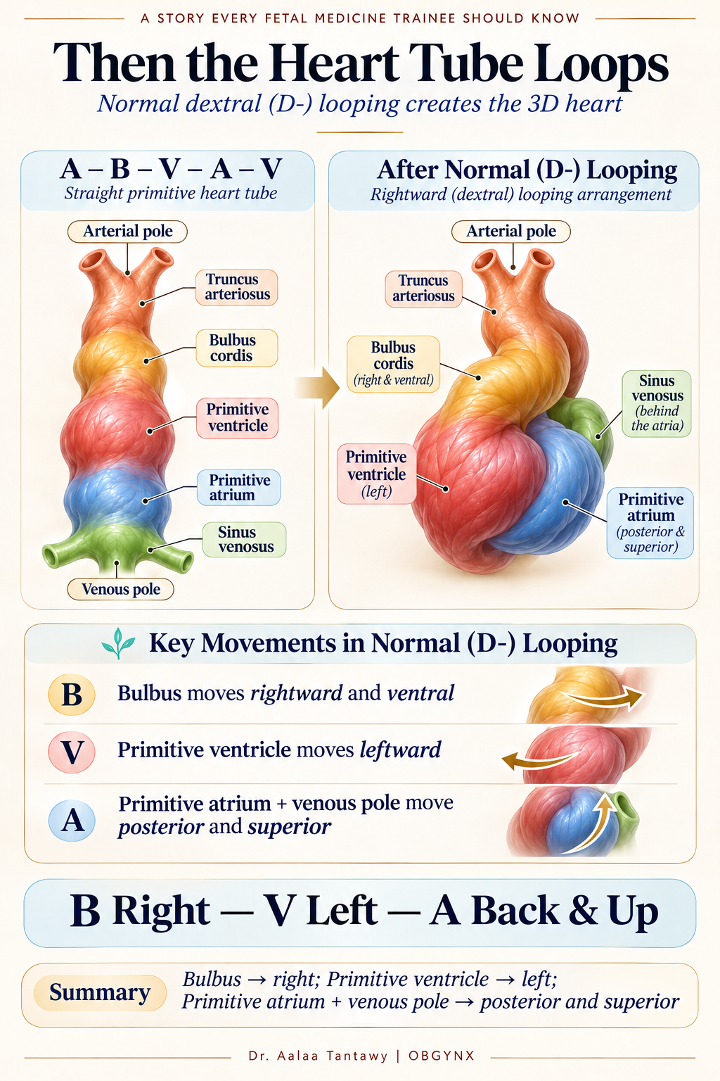
→ Shifts to the RIGHT
Places future right ventricle on the right side.
→ Shifts to the LEFT
Places future left ventricle on the left side.
→ Posterior & Superior
Swings up and behind the ventricles.
B Right — V Left — A Back & Up
The Final Memory Map
| Primitive Segment | Main Adult Derivative | Memory Key |
|---|---|---|
| A — Truncus arteriosus | Aorta + pulmonary trunk | A = Arteries |
| B — Bulbus cordis | Trabeculated RV + smooth outflow tracts of both ventricles | B = Right + BOTH exits |
| V — Primitive ventricle | Trabeculated LV | V = Left Ventricle |
| A — Primitive atrium | Trabeculated portions of both atria + auricles | A = Atria |
| V — Sinus venosus | Smooth RA (sinus venarum) + coronary sinus | V = Venous inflow |
The 10-Second Mnemonic & The Whole Story
Arteries
Right + BOTH exits
Left Ventricle
Atria
Venous inflow
• ABVAV starts the tube.
• The tube loops to the RIGHT.
• Arteriosus makes the great arteries.
• Bulbus makes the RV and both exits.
• Primitive ventricle makes the LV.
• Primitive atrium makes the trabeculated atria.
• Sinus venosus becomes the smooth RA and coronary sinus.
A-B-V-A-V → Arteries, Bulbus, Left Ventricle, Atria, Venous inflow.
Then: B Right — V Left — A Back & Up.
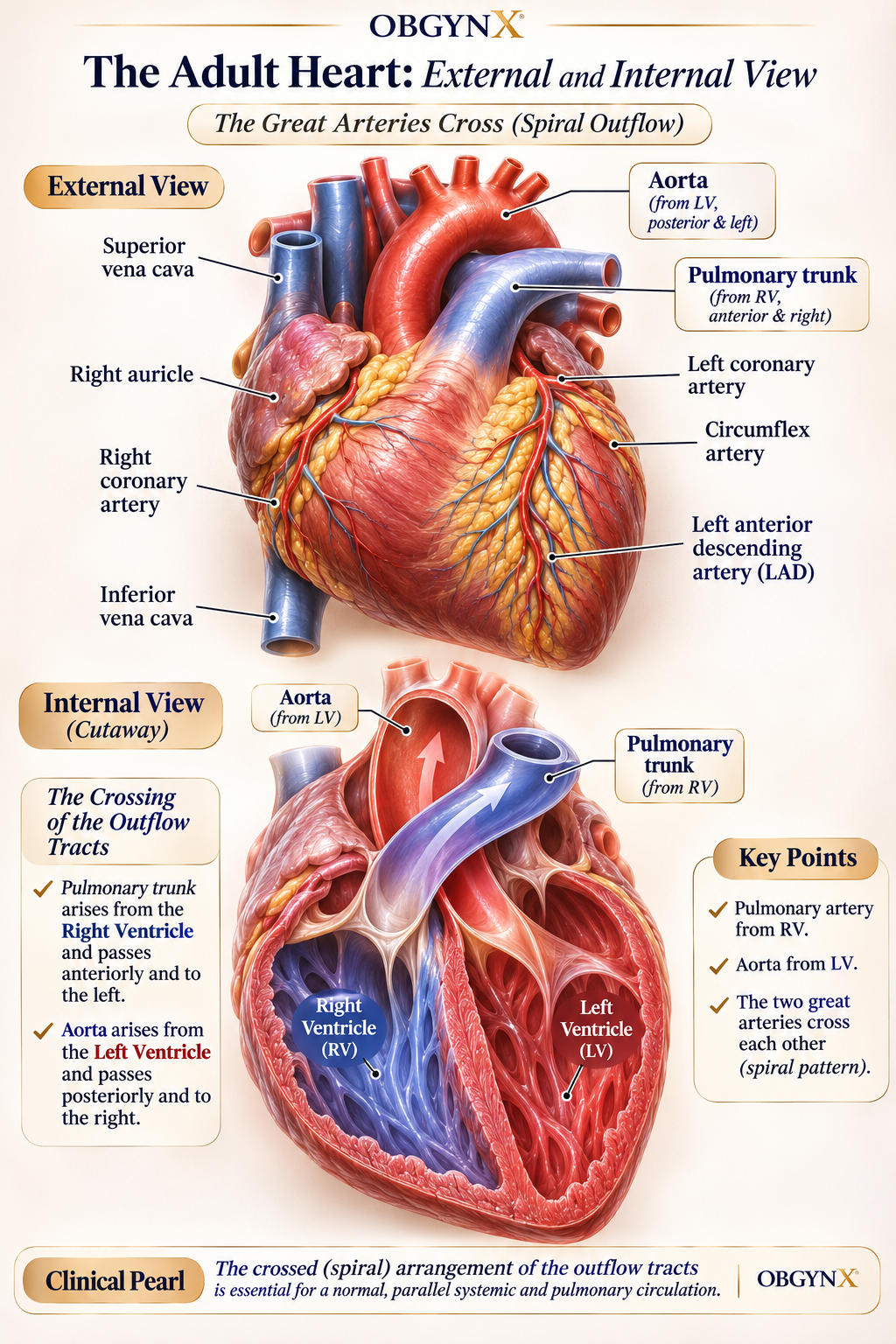
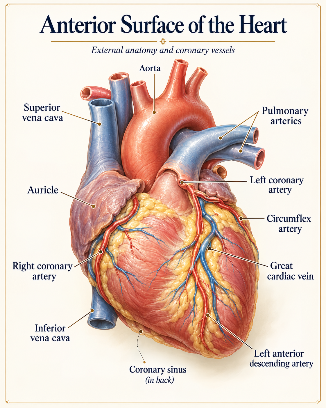
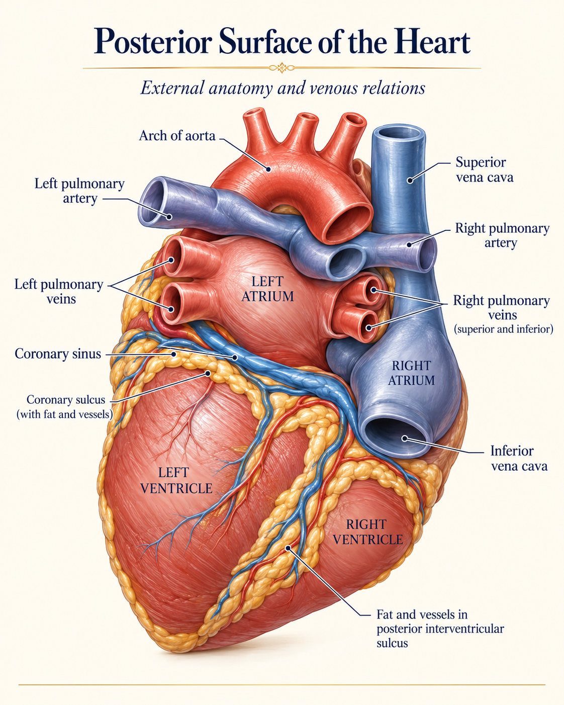
Adult Heart Anatomy & Spiral Outflow Crossing
How the 5 embryonic tube segments assemble into crossing great arteries and mature cardiac surfaces:
Detailed Course Syllabus
Intradecidual Sign & Gestational Sac
Early implantation milestones, mean sac diameter (MSD), and discrimination from ectopic fluid.
Yolk Sac & Embryonic Cardiac Pole
Appearance of the yolk sac, cardiac flicker detection via M-mode, and normal heart rates.
Rhombencephalon & Limb Bud Emergence
Normal physiologic cystic diamond of the hindbrain and early limb development.
Physiologic Gut Herniation & Early Cranium
Umbilical cord insertion, skull ossification, and distinguishing normal gut migration.
Learning Objectives
- Identify the double decidual sign and true gestational sac vs pseudogestational sac.
- Recognize the emergence of the secondary yolk sac at 5.5 weeks and evaluate normal morphology.
- Measure Crown-Rump Length (CRL) with precision from 6 to 11 weeks.
- Differentiate normal physiologic midgut herniation from true omphalocele between 8 and 11 weeks.
Clinical Relevance
Misinterpretation of normal early embryologic phenomena (such as physiologic midgut herniation or rhombencephalon) frequently leads to unnecessary termination counseling or patient distress.
Clinical Guidelines Aligned
- ISUOG Practice Guidelines: Performance of First-Trimester Fetal Ultrasound Scan
- Society of Radiologists in Ultrasound (SRU) Early Pregnancy Consensus
Explore Other Courses
Detailed First-Trimester Scan (12w 0d to 13w 6d)
Early anatomical survey, nuchal translucency caliper calibration, nasal bone assessment, and aneuploidy risk calculation.
Second and Third Trimesters (≥16+0 weeks)
Systematic screening of fetal structural anatomy, biometry measurements, and placental assessment.
Advanced Fetal Neurosonography
High-resolution multiplanar evaluation of the fetal central nervous system, midline structures, and posterior fossa.