The Primitive Heart Tube — The 5-Part Map
From the arterial pole to the venous pole: memorize the five primitive segments, the normal rightward D-loop, and their exact adult cardiac derivatives in one definitive framework.
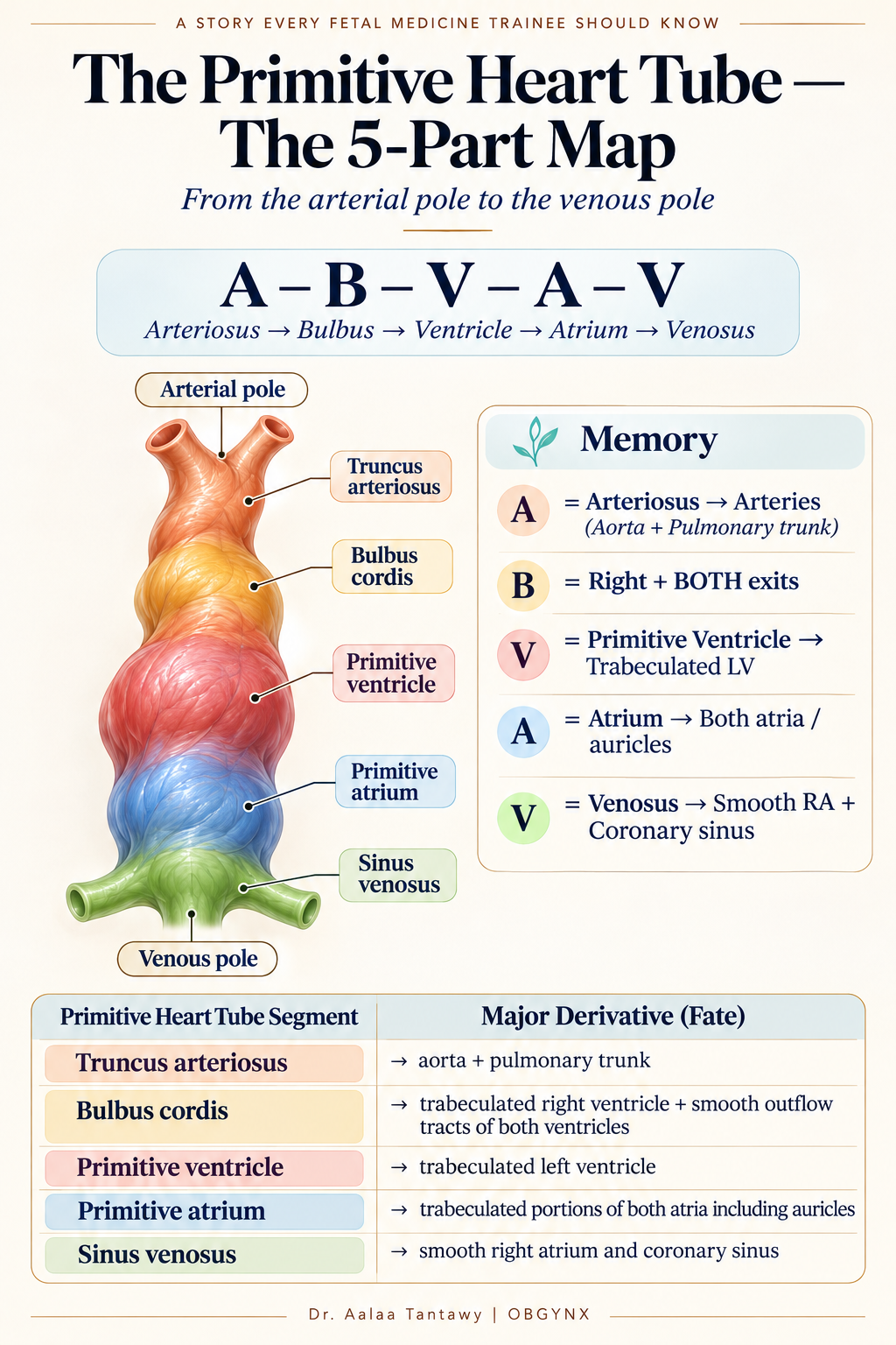
A – B – V – A – V (Arterial Pole to Venous Pole)
The primitive heart tube can be remembered from the cranial arterial outlet down to the caudal venous inflow as:
Truncus Arteriosus
The Future Great Arterial Outlet
Primary Adult Derivatives:
- →Aorta (Ascending)
- →Pulmonary Trunk
High-Yield Memory Rule:
A = Arteriosus → Arteries (Aorta + Pulmonary)
Failure of conotruncal septation leads to Persistent Truncus Arteriosus (single arterial trunk riding over a large VSD).
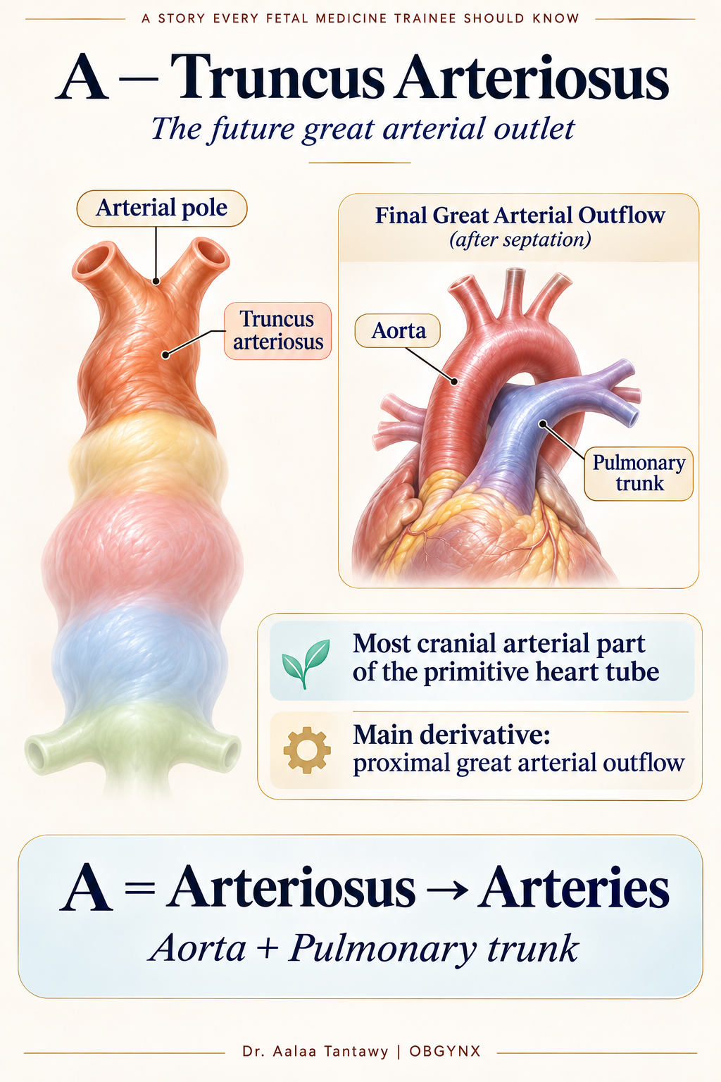 A — Truncus Arteriosus: Future Great Arterial Outflow (Aorta + Pulmonary Trunk)
The 5 Segments in Detail
Truncus Arteriosus — The Future Great Arterial Outlet
The truncus arteriosus is the most cranial arterial part of the primitive heart tube. It contributes to the proximal great arterial outflow:
• Aorta + Pulmonary trunk
A = Arteriosus → Arteries (Aorta + Pulmonary)
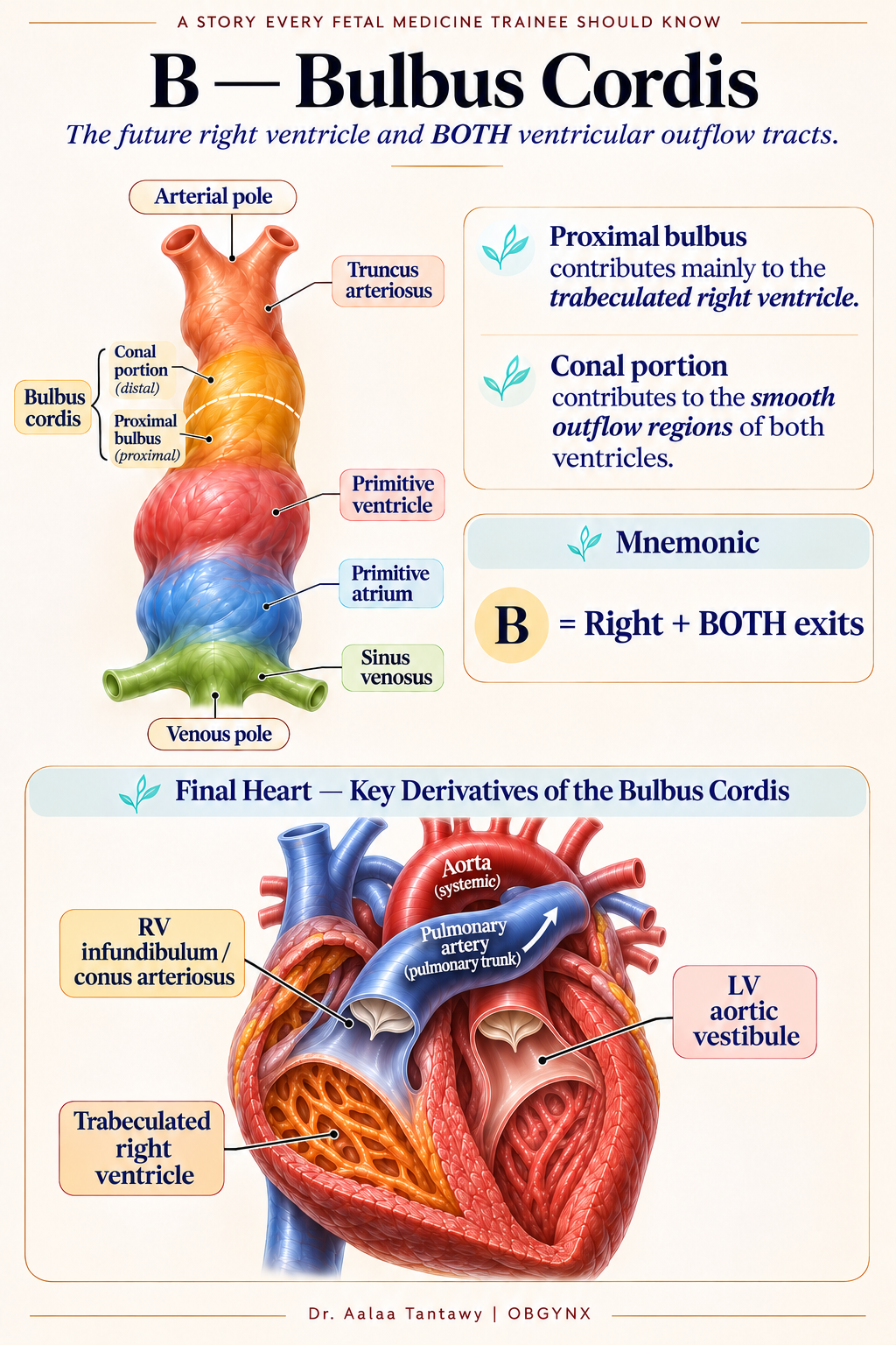
Bulbus Cordis — Future Right Ventricle & Outflow Tracts
The bulbus cordis has more than one important derivative across both sides of the heart:
• Proximal portion: Contributes mainly to the trabeculated right ventricle.
• Conal portion: Contributes to the smooth outflow regions of both ventricles:
- – RV infundibulum / conus arteriosus
- – LV aortic vestibule
B = Right + BOTH exits (Bulbus → RV + both ventricular outflow tracts)
This is the most useful way to remember it.
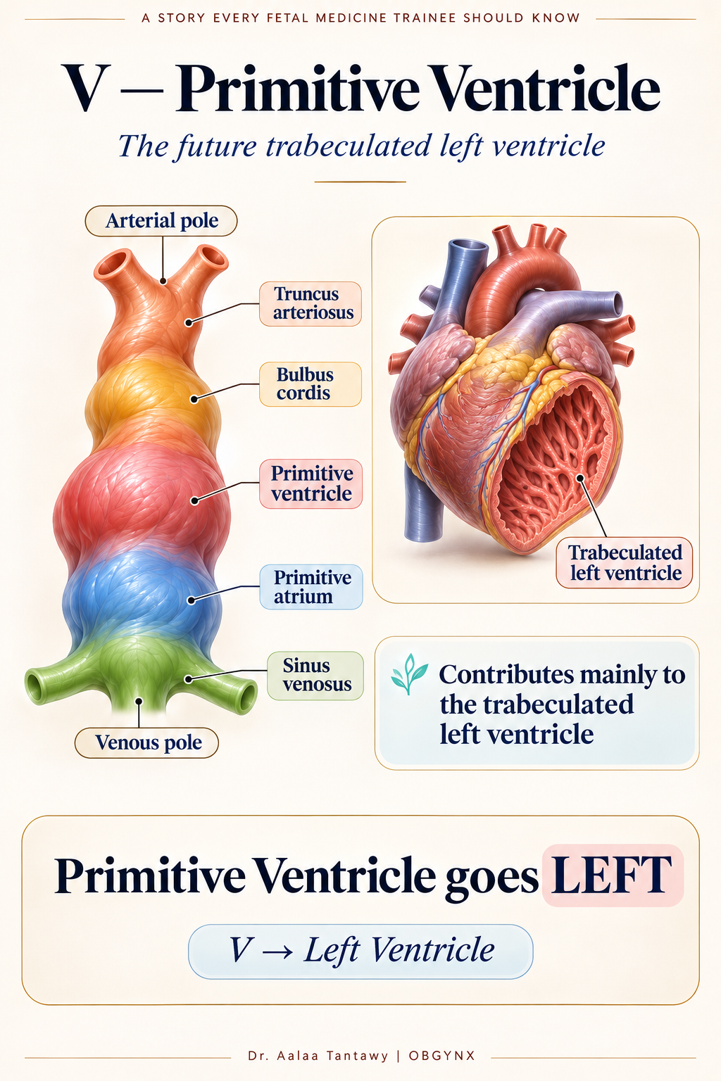
Primitive Ventricle — The Future Left Ventricle
The primitive ventricle contributes mainly to the:
• Trabeculated left ventricle
V → Left Ventricle
This is the odd one, so memorize it deliberately: Primitive Ventricle goes LEFT.
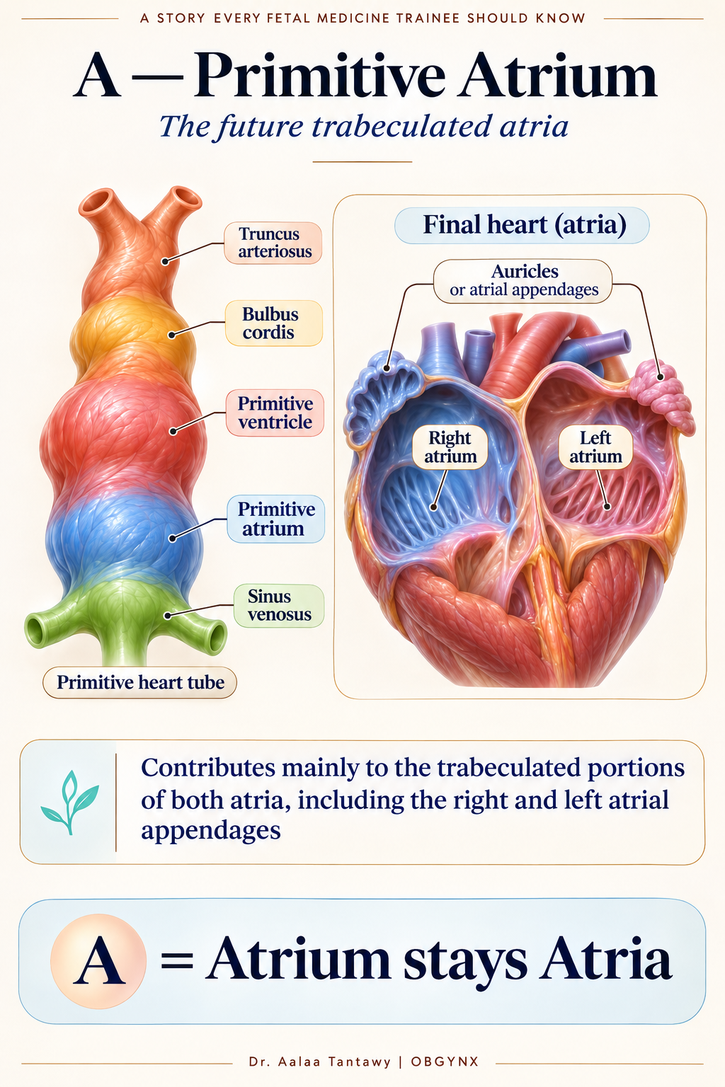
Primitive Atrium — The Future Trabeculated Atria
The primitive atrium contributes mainly to the:
• Trabeculated portions of both atria
including the Right and left atrial appendages / auricles (pectinate muscle walls).
A = Atrium stays Atria
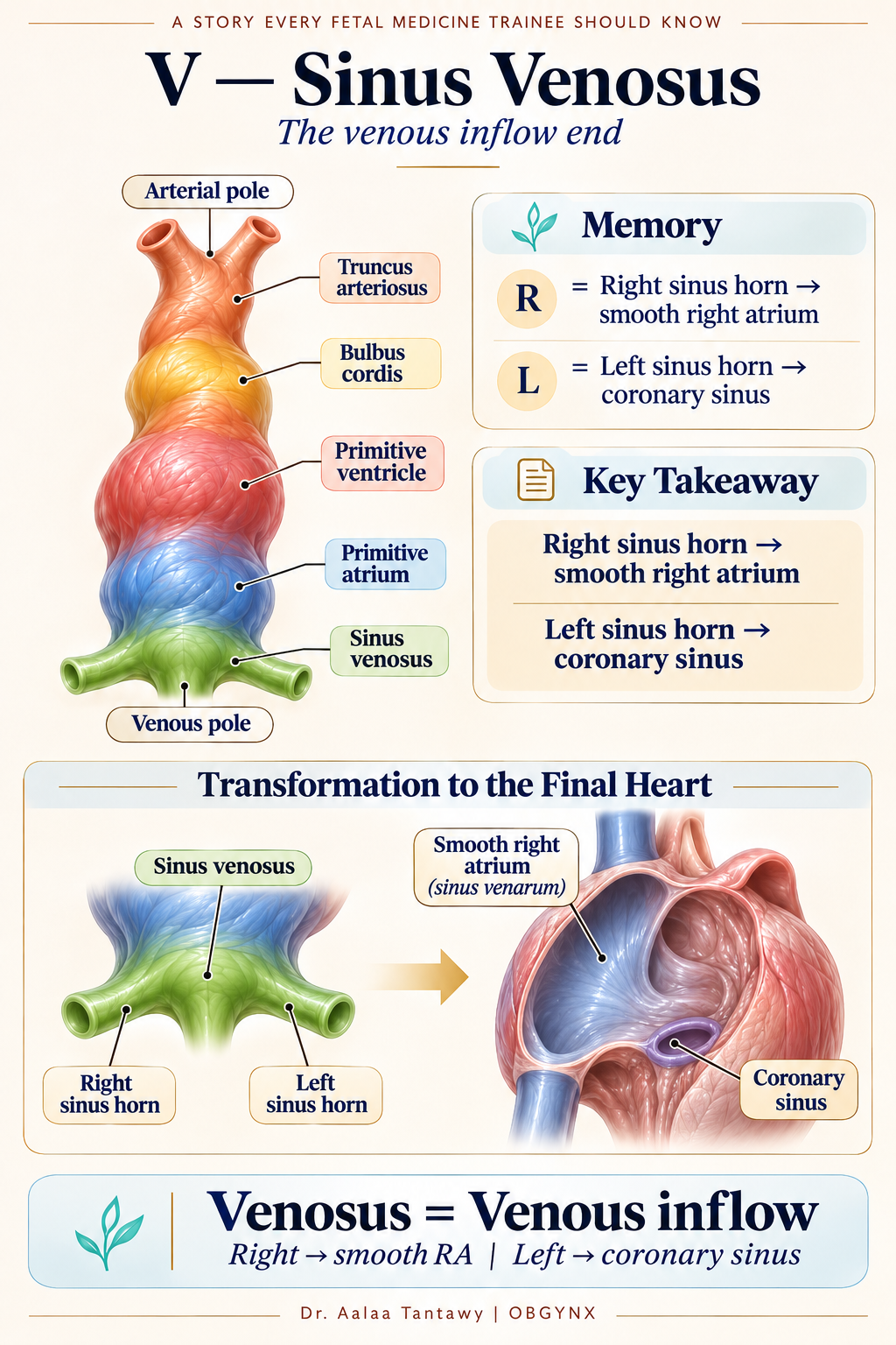
Sinus Venosus — The Venous Inflow End
The sinus venosus is the caudal venous end of the primitive heart. After looping and venous remodeling:
• Right sinus horn: Incorporated into the right atrium → forms the smooth-walled Sinus venarum.
• Left sinus horn: Mainly forms the Coronary sinus (drains heart veins).
Right → smooth Right Atrium • Left → Coronary sinus
Or simply: Venosus = Venous inflow.
Then the Heart Tube Loops (D-Looping)
The normal heart undergoes D-looping (Rightward looping / Dextral looping). This transforms a straight tube into a three-dimensional heart.
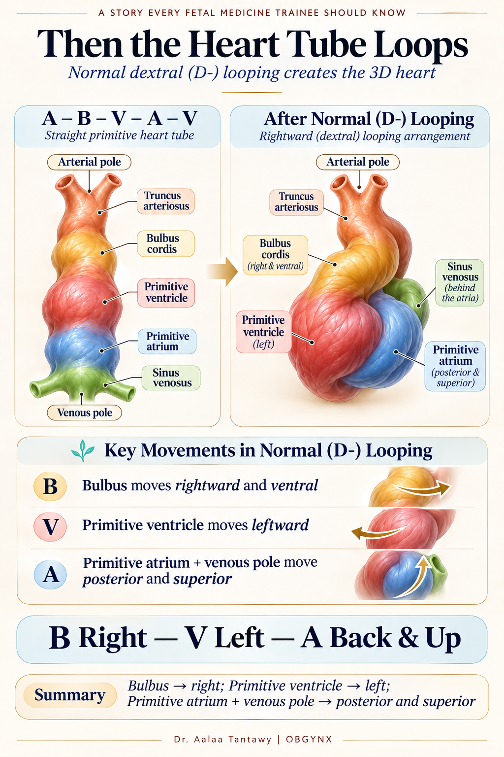
→ Shifts to the RIGHT
Positions future RV on the right side.
→ Shifts to the LEFT
Positions future LV on the left side.
→ Posterior & Superior
Swings behind and above the ventricles.
B Right — V Left — A Back & Up
The Final Memory Map
Start with A – B – V – A – V, then translate each letter into its major fate:
| Primitive Segment | Main Adult Derivative | Fast Recall Key |
|---|---|---|
| A —Truncus arteriosus | Aorta + pulmonary trunk | A = Arteries |
| B —Bulbus cordis | Trabeculated RV + smooth outflow tracts of both ventricles | B = Right + BOTH exits |
| V —Primitive ventricle | Trabeculated LV | V = Left Ventricle |
| A —Primitive atrium | Trabeculated portions of both atria + auricles | A = Atria |
| V —Sinus venosus | Smooth RA (sinus venarum) + coronary sinus | V = Venous inflow |
The 10-Second Mnemonic
Arteries
Right + BOTH exits
Left Ventricle
Atria
Venous inflow
And the whole story becomes:
• ABVAV starts the tube.
• The tube loops to the RIGHT.
• Arteriosus makes the great arteries.
• Bulbus makes the RV and both exits.
• Primitive ventricle makes the LV.
• Primitive atrium makes the trabeculated atria.
• Sinus venosus becomes the smooth RA and coronary sinus.
A-B-V-A-V → Arteries, Bulbus, Left Ventricle, Atria, Venous inflow.
Then: B Right — V Left — A Back & Up.
That is the primitive heart tube in one map.
Adult Heart Anatomy & The Great Outflow Crossing
See how the five primitive segments culminate in the definitive adult organ. The rightward D-looping and spiral conotruncal septation ensure the great arteries cross normally at right angles — the definitive hallmark examined on obstetric and fetal echocardiography scans.
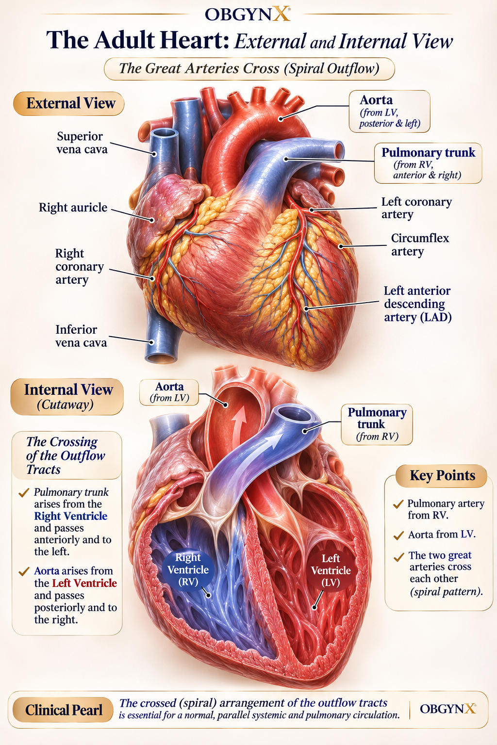
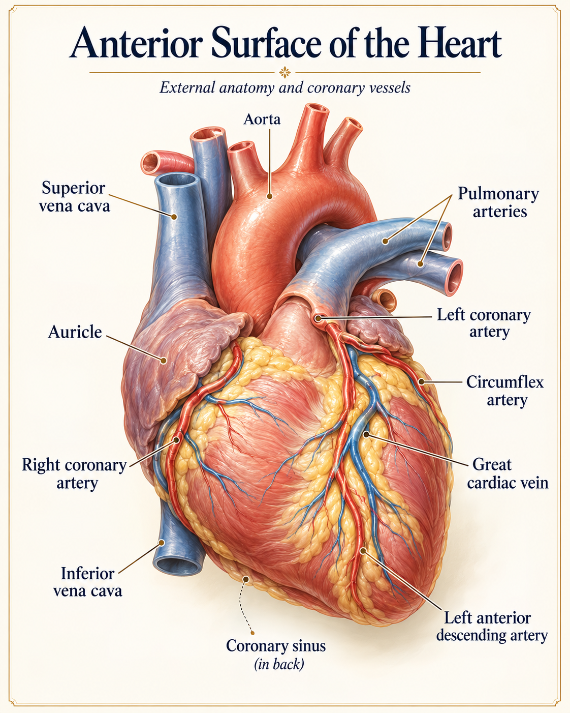
Demonstrates the dominant anterior position of the RV (formed from the bulbus cordis) and the crossing outflow tracts.
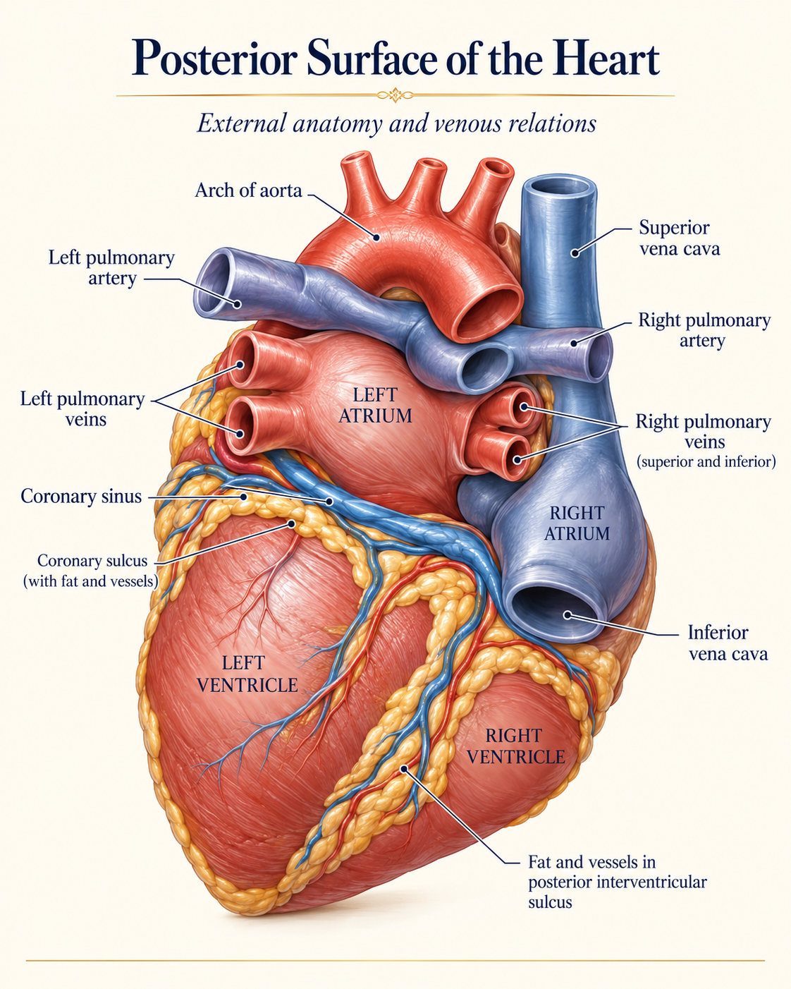
Shows the left atrial base receiving pulmonary veins and the coronary sinus running in the posterior atrioventricular groove.