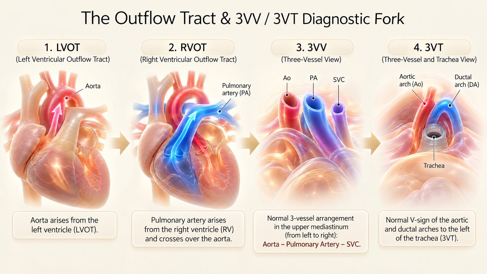
What is the outflow tract examination trying to prove?
A convincing fetal cardiac examination establishes arterial origin, course, branching, caliber, arch sidedness and blood-flow direction—not merely the presence of two moving vessels.
The right and left ventricular outflow tracts should be assessed as part of a continuous cephalad sweep from the four-chamber plane. First follow the vessel that arises from the morphologic left ventricle, then the vessel arising from the right ventricle. Continue cranially to evaluate vessel arrangement and then the aortic/ductal arch relationship to the trachea. The views overlap rather than representing rigid, isolated slices; cine loops are often more informative than individual still frames.
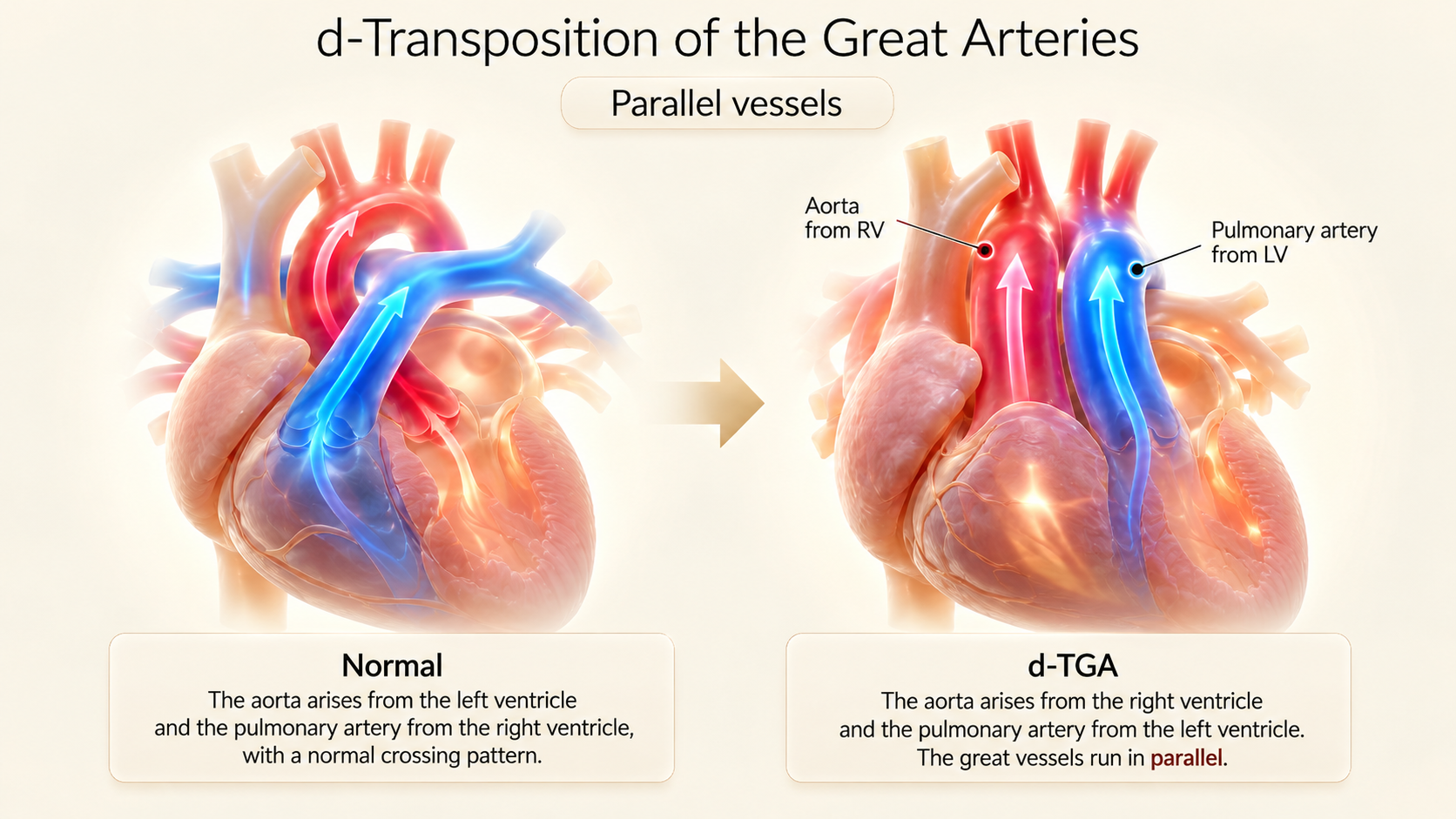
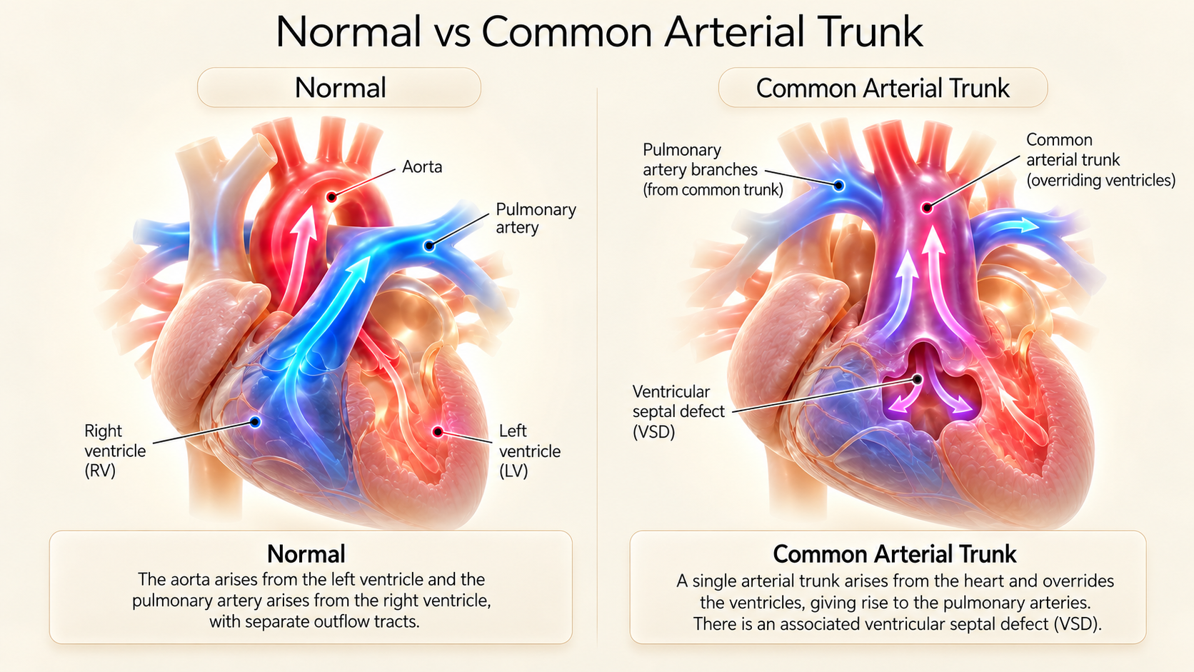
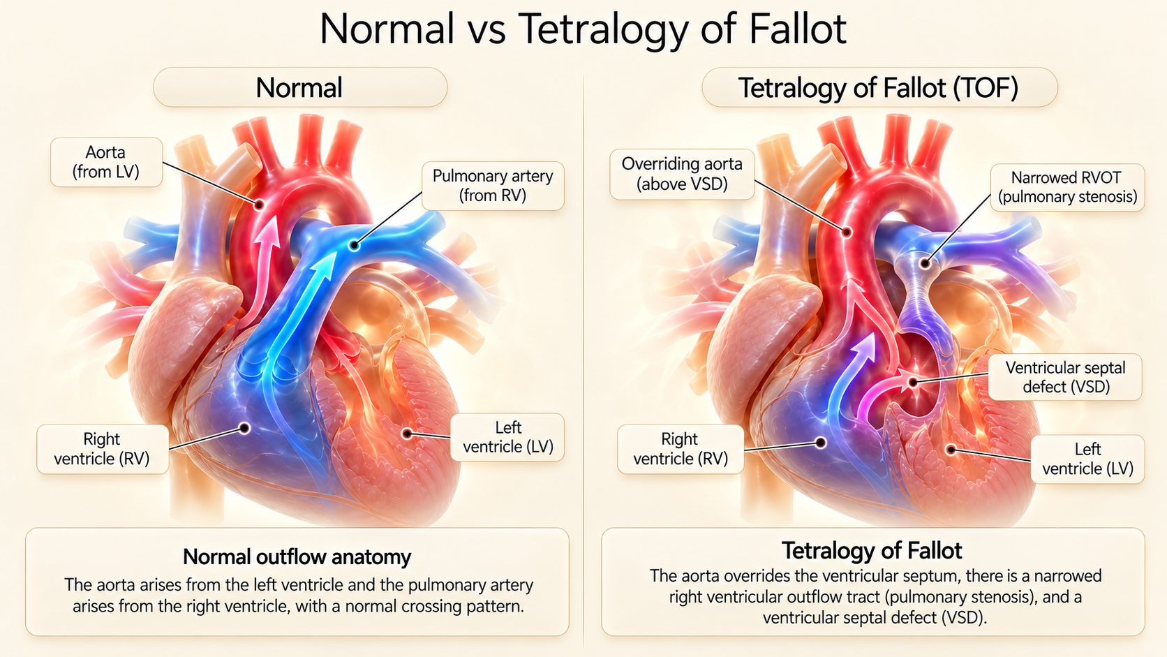
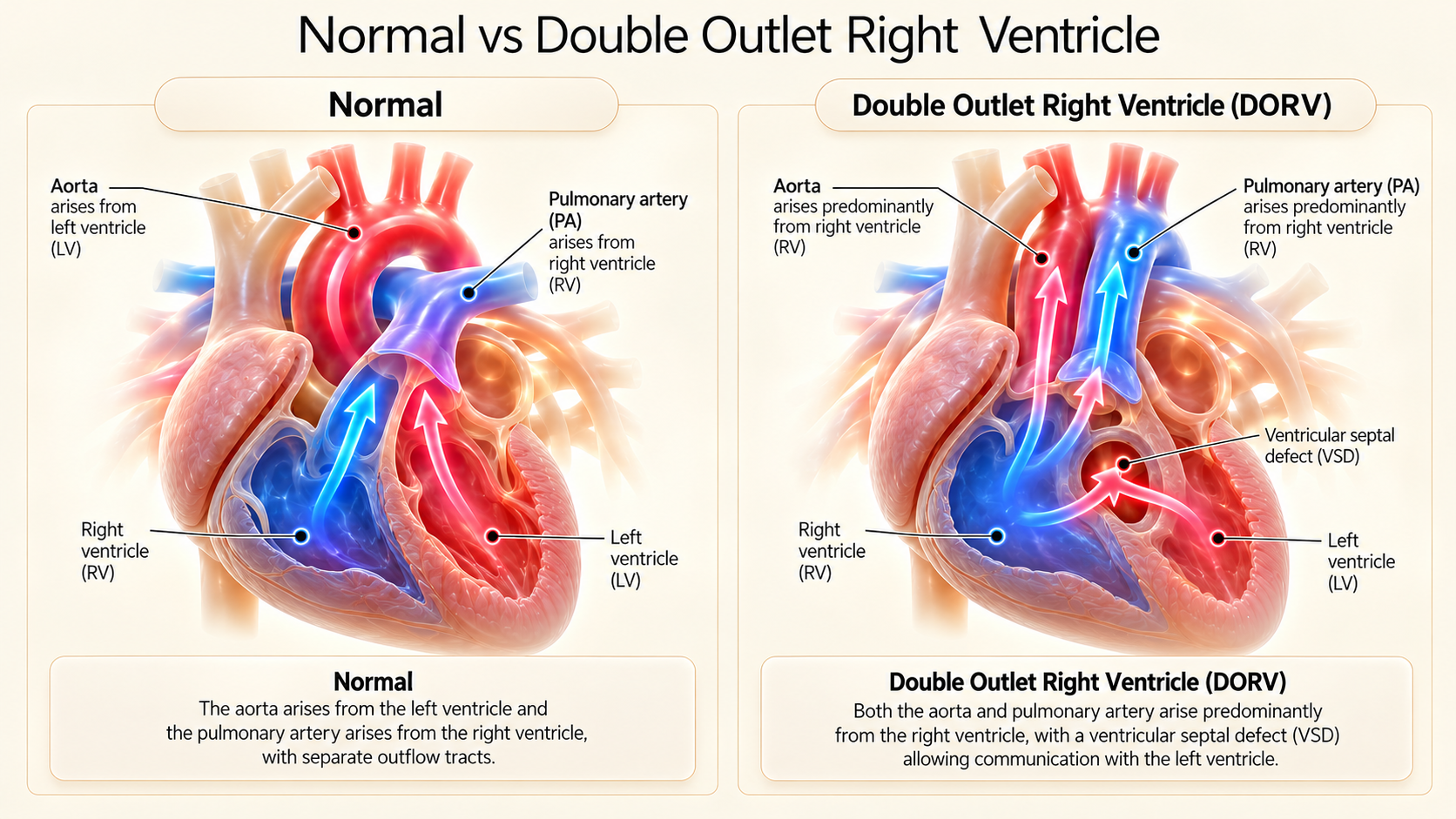
Normal outflow anatomy depends on ventriculoarterial concordance: left ventricle to aorta, right ventricle to pulmonary artery. The arteries usually cross close to their origin at approximately a 90-degree angle. The pulmonary artery subsequently bifurcates into branch pulmonary arteries (RPA and LPA), whereas the aorta can be followed into its arch, giving rise to the head and neck vessels.
"Every abnormal outflow image should lead to an anatomical question: Which artery is this, which ventricle does it leave, where does it go, and which anatomical landmark proves its identity?"
ISUOG guidelines recommend combining four-chamber, outflow and great-vessel views because major malformations—including d-transposition of the great arteries and tetralogy of Fallot—frequently present with a reassuring, symmetric four-chamber view. A normal 4CH view never replaces direct assessment of both great arteries.
Normal LVOT and RVOT: The Non-Negotiable Landmarks
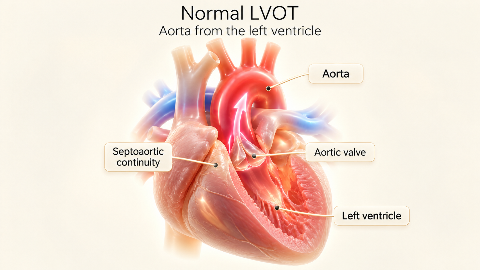
LVOT — What Must Be Demonstrated?
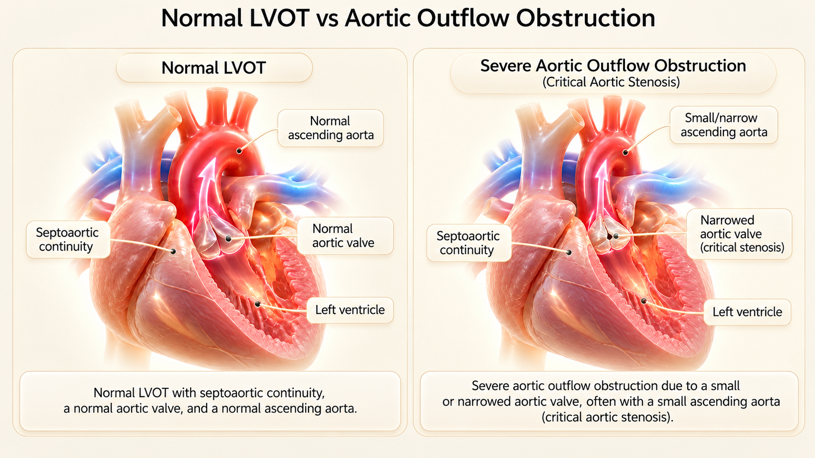
Left VentricleTrace the aortic root from the morphological LV. The ventricular septum must be continuous with the anterior wall of the proximal aorta (septo-aortic continuity). Assess the aortic valve’s appearance, opening, and follow the ascending aorta into its arch with head/neck branches.
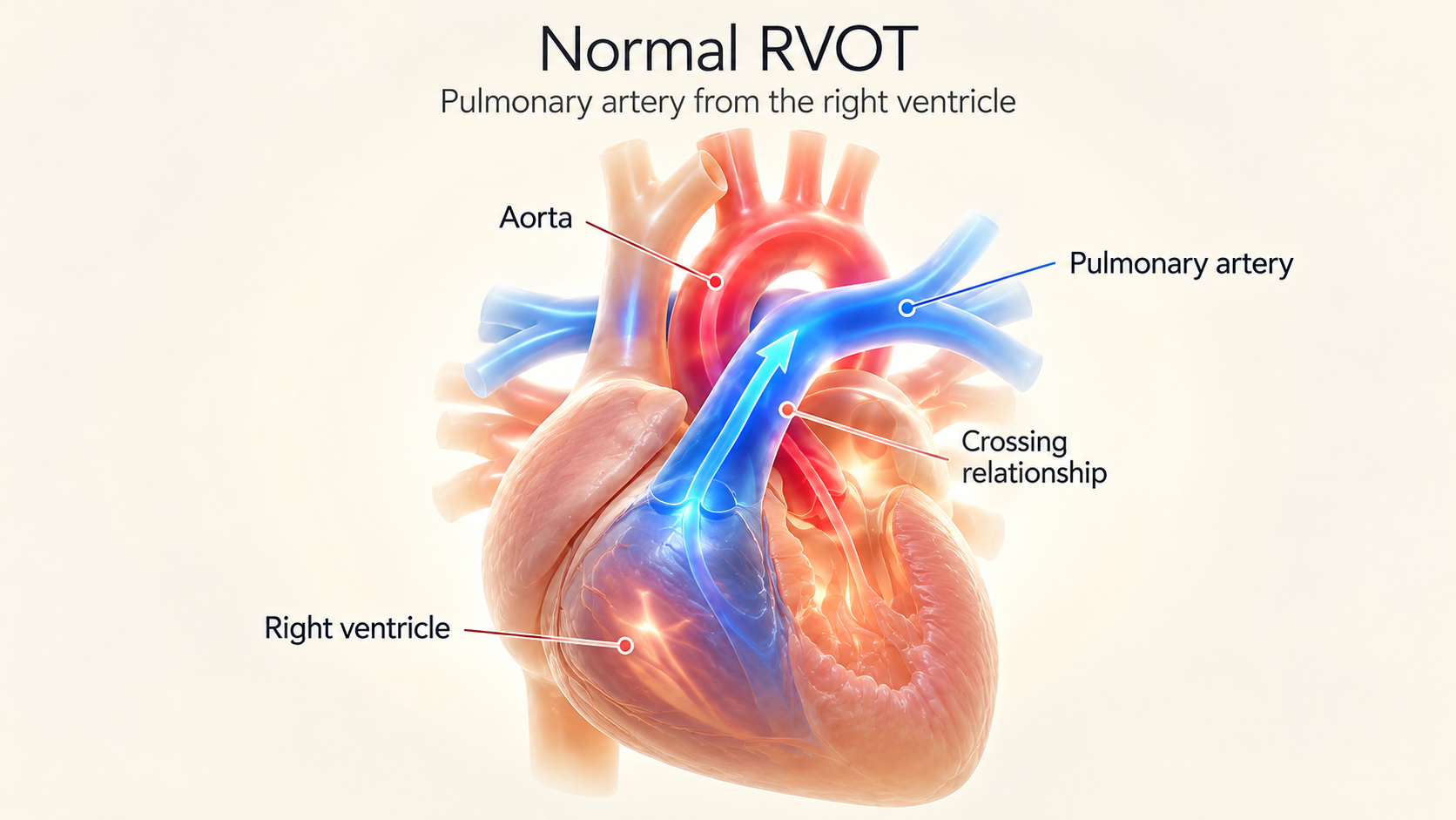
RVOT — What Must Be Demonstrated?
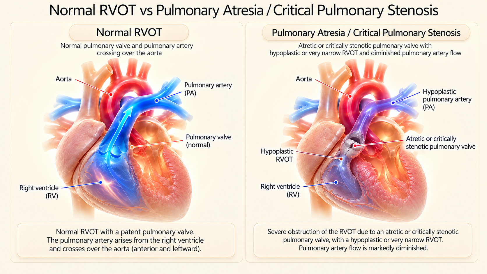
Right VentricleTrace the main pulmonary artery from the morphological RV (identified by muscular infundibulum and moderator band). Look for freely opening pulmonary leaflets and subsequent bifurcation into RPA and LPA. The pulmonary trunk crosses anteriorly over the aorta.
Understand the 3VV and the 3VT — They Answer Different Questions
Three-Vessel View (3VV)
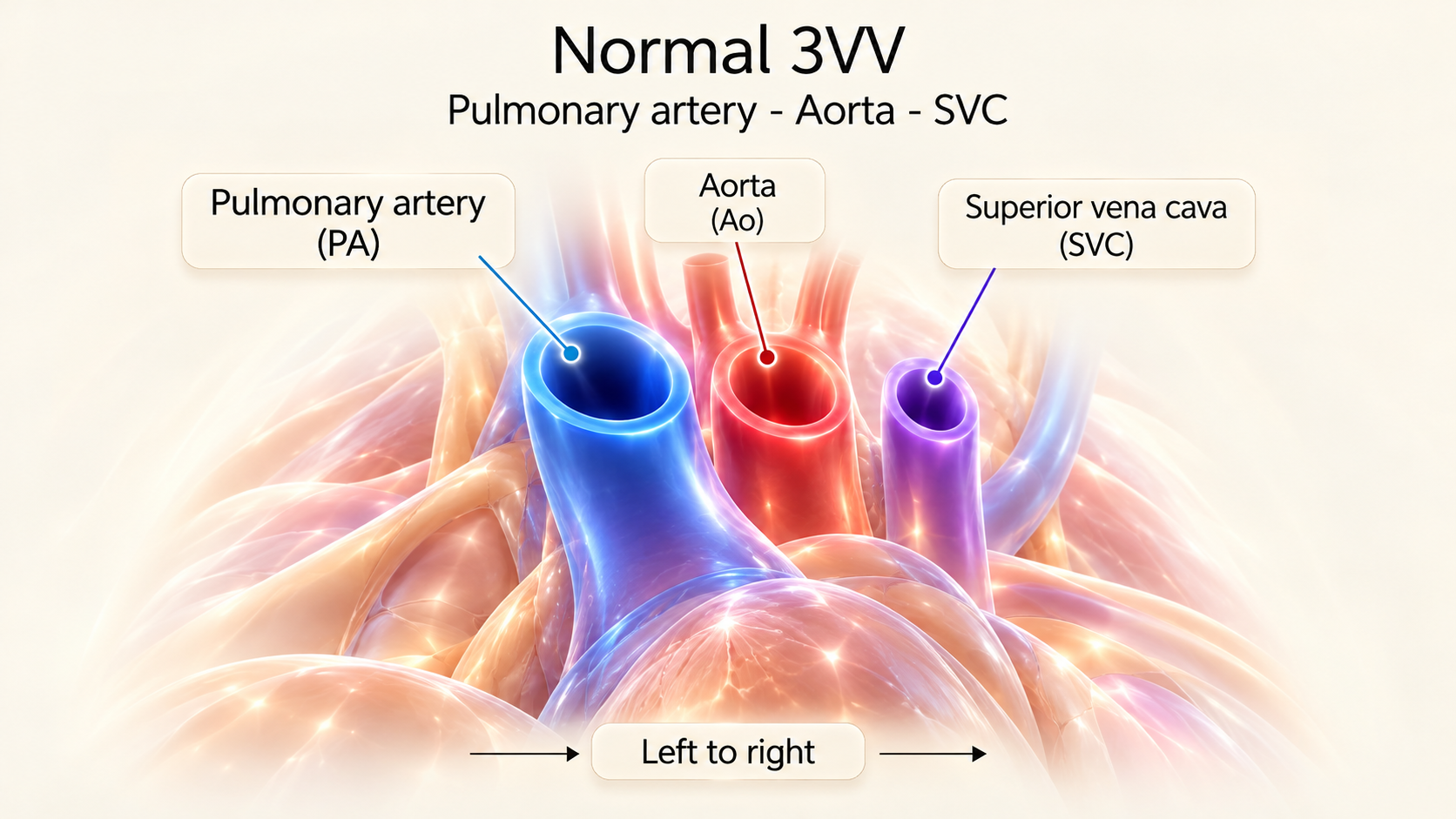
In a normal axial three-vessel view, the pulmonary artery is typically the most anterior and largest of the three vessels, followed by the ascending aorta, with the superior vena cava smaller and most posterior. The expected arrangement progresses obliquely from fetal left to fetal right. This view is therefore a rapid test of number, relative caliber, position and straight-line alignment.
Three-Vessel-and-Trachea View (3VT)
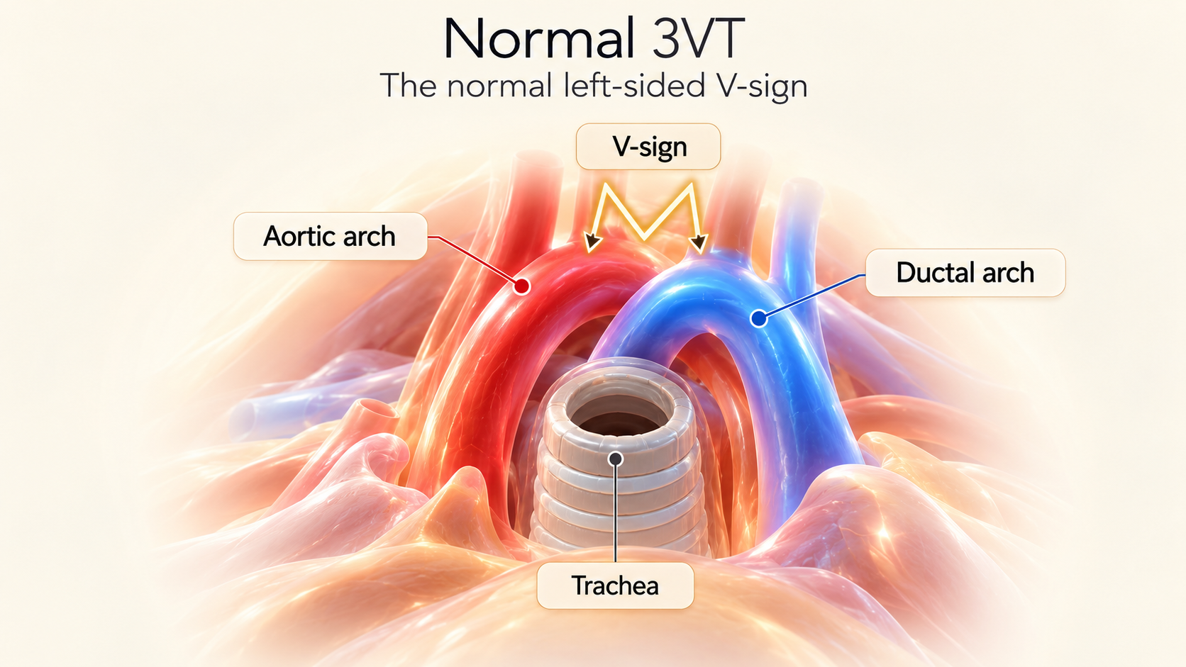
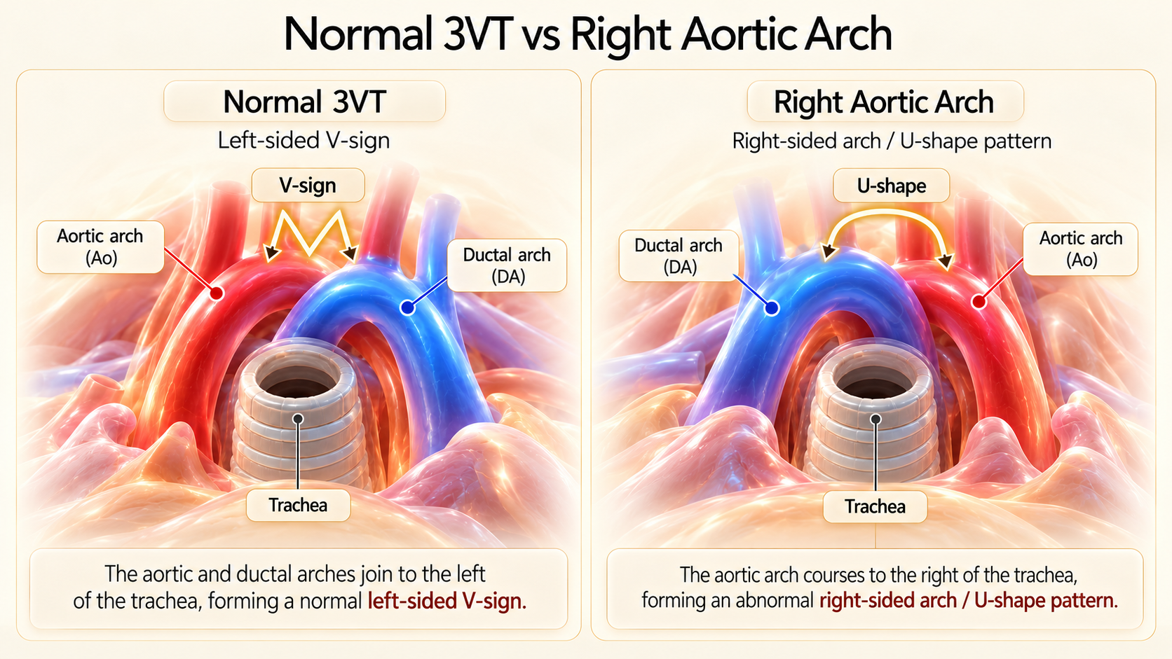
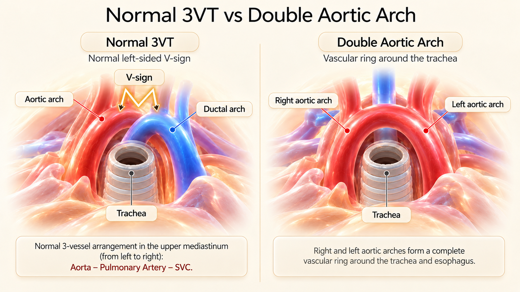
With a further cephalad sweep and slight transducer tilt, identify the aortic arch, the ductal arch, the superior vena cava, and the fluid/air-filled trachea. In typical left-sided arch anatomy, the two arches course to the left of the trachea and join the descending aorta in the characteristic acute V pattern.
When LVOT or RVOT Is Abnormal: Classify Before Naming a Disease
Start by confirming atrioventricular and ventriculoarterial connections. If the vessel connected to the RV appears to be the aorta, and the vessel connected to the LV appears to be the pulmonary artery, consider d-transposition of the great arteries (d-TGA). The proximal great vessels run in parallel rather than crossing at 90 degrees. An elongated or I-shaped anterior aorta on the upper mediastinal sweep is a helpful clue, not a standalone diagnostic proof.
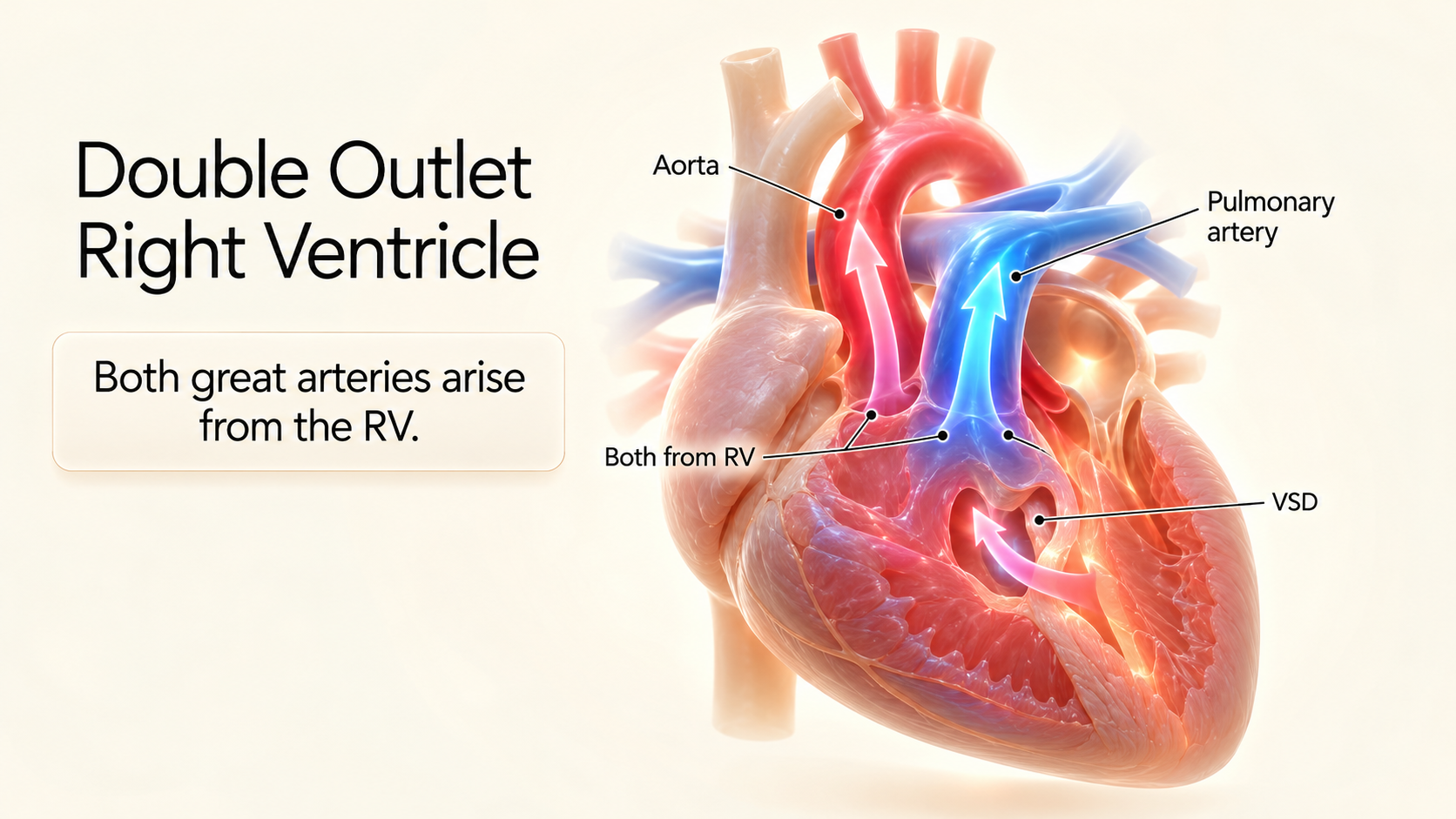
If both great arteries predominantly connect to the morphological RV, consider double-outlet right ventricle (DORV). Determine VSD location (subaortic vs. subpulmonary / Taussig-Bing), arterial relationship, and presence of subpulmonary stenosis.
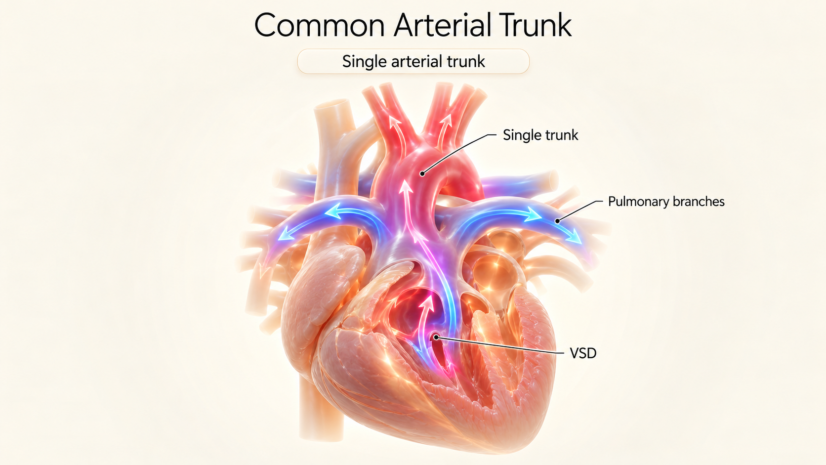
Do not equate "I can see only one vessel" with a final diagnosis. First exclude poor acoustic windows, extreme arterial hypoplasia, or an atretic outlet. If one common arterial trunk supplies systemic, pulmonary, and coronary circulations over an outlet VSD, consider common arterial trunk (persistent truncus arteriosus).
In pulmonary atresia with VSD (pseudotruncus), the pulmonary arteries are supplied retrograde through the ductus arteriosus or major aortopulmonary collateral arteries (MAPCAs), with no antegrade flow from the RV.
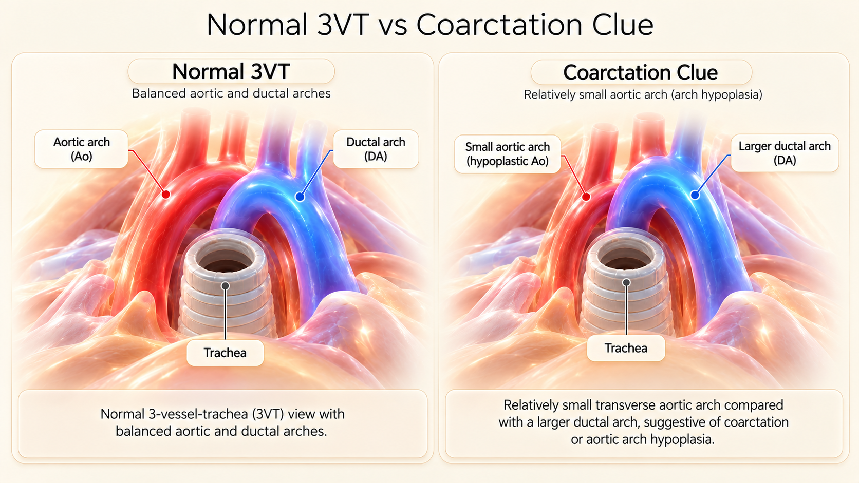
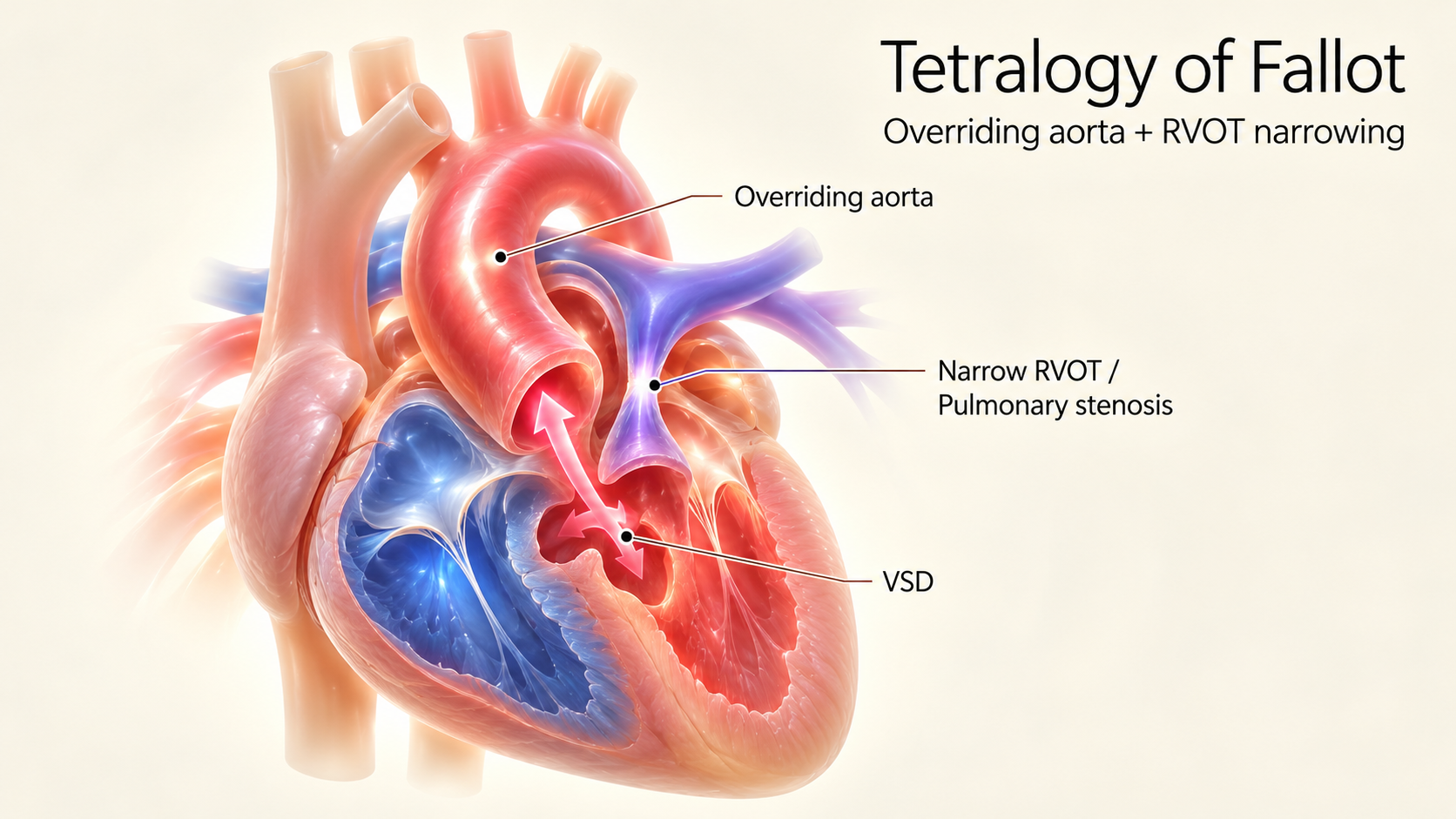
A small pulmonary artery, reduced RVOT caliber, or restricted pulmonary leaflet opening raises concern for pulmonary stenosis, pulmonary atresia, and the TOF spectrum. Conversely, a small ascending or transverse aorta, especially relative to a dilated ductal arch, raises concern for left-sided outflow obstruction, arch hypoplasia, or coarctation of the aorta.
Six Abnormal 3VV/3VT Patterns You Should Never Ignore
| Pattern | Important Differential Diagnosis | What Must Be Checked Next? |
|---|---|---|
| 1. PA unusually small relative to Ao | TOF spectrum; significant pulmonary stenosis/atresia; selected complex outflow defects. | RVOT connection, pulmonary valve opening, MPA & branch PAs, outlet VSD, ductal flow direction. |
| 2. Ao/arch unusually small relative to ductal arch | Arch hypoplasia/coarctation; critical aortic stenosis; left-heart obstruction spectrum. | LVOT, aortic valve, transverse arch & isthmus, flow direction, ventricular sizes, sagittal arch view. |
| 3. Arterial vessels do not cross as expected | d-TGA; DORV; other complex ventriculoarterial discordance. | Trace origins from each ventricle, confirm branching (PA bifurcates vs. Ao head/neck), assess VSD. |
| 4. Apparently single dominant arterial channel | Common arterial trunk; pulmonary atresia with VSD; severe arterial hypoplasia; acoustic shadow. | Identify pulmonary arteries and their source; look for common truncal valve and outlet VSD. |
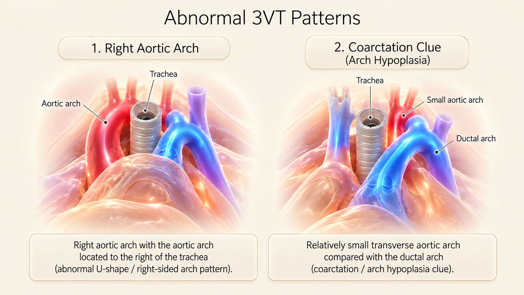
| 5. Abnormal arch position around trachea | Right aortic arch, double aortic arch, vascular ring, and branching variants (ARSA). | Confirm aortic arch side relative to trachea, ductal sidedness, arch branching, and U-pattern. |
| 6. Apparent additional mediastinal vessel | Persistent left SVC (PLSVC); dilated azygos/hemiazygos; other systemic venous variants. | Follow venous course caudally, inspect coronary sinus for dilation, assess systemic venous return. |
Turn the Abnormal View into a Focused Differential Diagnosis
Tetralogy of Fallot (TOF)Conotruncal
Look for an outlet/malalignment VSD with the aorta overriding the septum and varying degrees of RV outflow obstruction. The pulmonary artery is characteristically small. The 4CH view can appear entirely normal. Assess pulmonary valve, MPA, branch PAs, ductal flow, and aortic arch sidedness (~25% right arch).
d-Transposition (d-TGA)Discordant
The aorta arises from the RV and the pulmonary trunk arises from the LV, running parallel. The 4CH view is commonly completely normal. Demonstrate both ventricular origins rather than relying on a single 'I-sign'. Early prenatal detection allows planned prostaglandin infusion and urgent balloon atrial septostomy.
Double-Outlet Right Ventricle (DORV)Complex RVOT
Both great arteries arise predominantly (>50%) from the morphological RV. Characterize the VSD relationship to the semilunar valves (subaortic vs. subpulmonary / Taussig-Bing anomaly) and evaluate for pulmonary stenosis or coarctation. Subtypes have vastly different postnatal surgical pathways.
Common Arterial Trunk (Truncus)Single Trunk
A single arterial trunk leaves the heart over an outlet VSD, giving rise to coronary, systemic, and pulmonary arteries. Search for pulmonary branch origins from the posterior/lateral aspect of the common trunk, evaluate the dysplastic truncal valve, and rule out 22q11.2 microdeletion.
Pulmonary Atresia / Critical PSDuct-Dependent
The pulmonary valve shows restricted opening or complete imperforation. Color Doppler demonstrates aliasing turbulence across a stenotic valve or retrograde aorta-to-pulmonary flow through the ductus arteriosus in ductal-dependent pulmonary circulation.
Coarctation / Arch HypoplasiaArch Defect
Marked caliber narrowing of the transverse aortic arch and isthmus relative to the ductal arch, accompanied by right-heart dominance (RV > LV, PA > Ao). Prenatal diagnosis remains challenging because the ductus arteriosus carries right-to-left flow, masking postnatal ductal closure stenosis.
The Trachea Is Your Orientation Landmark
When the normal left-sided V-pattern changes, stop and establish the anatomy in an ordered fashion. Identify the trachea (hyperechoic cartilage ring surrounding an echo-free lumen). Find the aortic arch and state whether it courses to the left or right of the trachea. Then identify the ductal arch and its sidedness. Finally trace the descending aorta and head/neck vessel pattern where possible.
Color Doppler Reveals Direction—If You Interpret It Correctly
Color Doppler helps confirm vessel patency, laminar versus disturbed flow, and unusual direction relative to anatomy. It should be used with appropriate scale, gain, and beam angle, accounting for fetal movement and aliasing. Red and blue do not mean oxygenated and deoxygenated blood. They represent direction relative to the transducer according to the displayed color map, which may be inverted.
Follow flow through the pulmonary artery and ductus, and through the ascending and transverse aorta. In suspected severe left-sided obstruction, retrograde flow within the transverse arch or isthmus can be a crucial finding. In suspected critical pulmonary outflow obstruction, duct-dependent pulmonary supply is evidenced by aorta-to-pulmonary retrograde ductal flow.
The 7 Most Common Outflow Pitfalls
- Using an oblique imaging plane and misidentifying an artery.
- Assuming two arteries cross without systematically following their ventricular origins.
- Calling an apparent extra vessel a persistent left SVC without tracing venous continuity into the coronary sinus.
- Calling coarctation of the aorta based on ventricular or arch size disproportion alone without multiplanar confirmation.
- Using a color map as a surrogate for anatomical oxygenation rather than transducer vector orientation.
- Making a major surgical diagnosis from a single still frame rather than a carefully recorded high-frame-rate cine sweep.
- Confusing the hockey-stick ductal arch with the candy-cane aortic arch on a longitudinal sagittal image.
OBGYNx Interactive Outflow Diagnostic Fork
Choose the primary observed outflow abnormality. The interactive algorithm demonstrates the next logical anatomical questions, discriminating views, and major pitfalls.
Abnormal Great-Artery Crossing
Important possibilities: Complete Transposition of Great Arteries (d-TGA), Double-Outlet Right Ventricle (DORV), Congenitally Corrected TGA (cc-TGA), and complex ventriculoarterial discordance.
Trace each vessel back to its morphological ventricle. Confirm pulmonary bifurcation into RPA/LPA versus aortic arch giving head and neck branches. Search for outlet VSD and infundibular obstruction.
Three Clinical Interpretation Exercises
Normal 4CH, Unusual Parallel Arteries
You demonstrate an artery leaving the morphologic RV that curves into an arch with head/neck vessels, and a separate pulmonary artery arising from the morphologic LV that bifurcates. What does this establish?
Small PA, Overriding Aorta
An outlet VSD is identified with a malaligned overriding aorta and a diminutive pulmonary outflow. What is the key diagnostic spectrum?
Small Transverse Aortic Arch
The 3VT view reveals a much narrower aortic arch limb than ductal arch limb, with mild ventricular disproportion (RV > LV). Can you definitively diagnose coarctation of the aorta?
Note: These represent structured clinical reasoning scenarios for medical education, not individual patient case reports.
Test Your Outflow Examination Principles
8 high-yield clinical reasoning questions. Answer each to receive immediate validation and detailed explanations.
The Seven-Item Outflow Practical Checklist
- Confirm situs and morphological ventricular identity: Match stomach to left side, apex to left side, moderator band to RV, smooth septum to LV.
- Trace LVOT to the aortic arch: Establish septo-aortic continuity, verify aortic leaflet opening, and follow the transverse arch into head and neck vessels.
- Trace RVOT to the pulmonary artery: Demonstrate main pulmonary trunk arising from RV infundibulum and bifurcating into RPA and LPA.
- Verify normal 90-degree crossing: Confirm that the RVOT crosses anteriorly over the LVOT; rule out parallel ("I-sign") arterial courses.
- Record 3VV caliber rule: Verify that PA > Ao > SVC and that the three vessels align along a straight diagonal line.
- Use 3VT to assess arch convergence and trachea: Confirm that the aortic arch and ductal arch converge to the LEFT of the trachea in an acute V-sign.
- Document any abnormal finding with cine sweeps: Never rely on a single still image. Assess color Doppler flow direction and refer for specialist fetal echocardiography.
References and Medical Review
- Carvalho JS, et al. ISUOG Practice Guidelines (updated): fetal cardiac screening. Ultrasound Obstet Gynecol. 2023;61:788–803.
- Moon-Grady AJ, et al. Guidelines and Recommendations for Performance of the Fetal Echocardiogram. American Society of Echocardiography, 2023.
- Anton T, et al. The Fetal 3-Vessel Views: An Illustrative Case-Based Tutorial. J Ultrasound Med. 2019.
- ISUOG VISUOG: Right Aortic Arch.
- ISUOG VISUOG: Coarctation of the Aorta.
- Prenatal diagnosis of transposition of the great arteries: an updated review. 2020.