Abnormal Fetal Four-Chamber View: A Step-by-Step Diagnostic Approach
From the First Abnormal Ultrasound Finding to Congenital Heart Defect Differential Diagnosis
Section 1 — Why the Four-Chamber View Matters
For more than four decades, the fetal four-chamber view (4CV) has stood as the universal baseline for prenatal cardiac screening. First incorporated into standard obstetric protocols in the late 1980s, obtaining a normal 4CV excludes over 50% of major congenital heart defects (CHD)—including complete atrioventricular septal defects (cAVSD), hypoplastic left heart syndrome (HLHS), severe tricuspid atresia, and Ebstein anomaly.
- Chamber Balance: Two symmetric ventricles and two symmetric atria without severe hypoplasia.
- Normal Cardiac Crux: Intact lower interatrial and upper interventricular septa with normal 1–2 mm apical offset of the tricuspid valve.
- Normal Cardiac Axis: Apex directed at 45° ± 20° to the left, ruling out gross intrathoracic mass displacement or dextrocardia.
- Normal Size: Cardiac area occupying ~1/3 of the chest, excluding gross cardiomegaly or pulmonary hypoplasia.
- Conotruncal Malformations: Complete D-TGA, Tetralogy of Fallot, Truncus Arteriosus, and Double Outlet Right Ventricle frequently have a completely normal 4CV!
- Aortic Arch Obstruction: Coarctation of the aorta often presents with subtle or no chamber asymmetry in mid-gestation.
- Semilunar Valve Lesions: Mild-to-moderate pulmonary stenosis and bicuspid aortic valve do not alter four-chamber balance.
- Total Anomalous Pulmonary Venous Connection (TAPVC): Chambers can appear deceptively balanced without close venous Doppler interrogation.
Normal vs Abnormal Four-Chamber View Benchmark
Side-by-side comparative anatomical graphic illustrating normal 4CV criteria (1:1 chamber symmetry, 45° axis, differential offset at cardiac crux, intact IVS/IAS) juxtaposed with three primary abnormal archetypes: (1) Ventricular Disproportion (HLHS/CoA), (2) Crux Dissolution (cAVSD), and (3) Extreme Right Atrial Dilation (Ebstein Anomaly).
Normal Four-Chamber View (4CV) Architectural Criteria
The Gold Standard Transverse Plane of the Fetal Heart
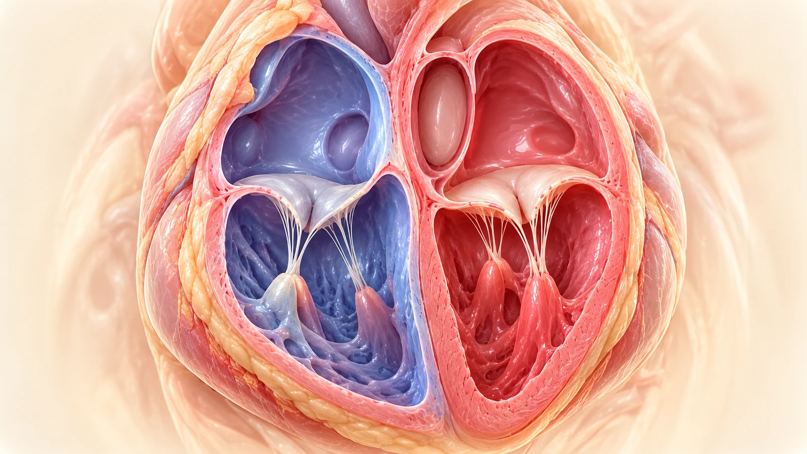
The normal transverse four-chamber view demonstrates symmetrical left and right ventricular chambers (ratio 1:1), intact ventricular and atrial septa, patent foramen ovale flap bulging exclusively into the left atrium, normal crux offset where the tricuspid valve septal leaflet inserts 1–2 mm more apical than the mitral valve, and at least two pulmonary veins draining into the smooth posterior left atrium.
- Ventricular symmetry: LV and RV equal in length and transverse dimension (ratio 0.95–1.05)
- Cardiac crux offset: Tricuspid valve inserts 1–2 mm closer to the apex than the mitral valve
- Interatrial septum: Flap of fossa ovalis oscillates exclusively into the left atrium
- Pulmonary venous connection: Minimum two pulmonary veins visualized entering the smooth posterior left atrium
Section 2 — First Confirm the View Is Technically Correct
Before pronouncing a four-chamber view abnormal, the sonographer must systematically audit technical quality. Up to 40% of suspected cardiac abnormalities referred to specialist centers represent artifactual distortions caused by improper plane angulation, rib shadowing, or acoustic drop-out.
Determine fetal lie (cephalic vs breech) and spine orientation before identifying left and right. Never assume left or right based on probe orientation alone.
Verify a complete, single rib shadow on each lateral thoracic wall. An oblique cut showing multiple ribs artificially foreshortens the ventricles, mimicking disproportion or a false VSD.
The fetal heart must occupy at least 1/3 to 1/2 of the ultrasound display monitor. Scanning on full-body presets obscures valve leaflets and crux anatomy.
- Coronal/Oblique Tilt: Tilting the probe towards the fetal neck brings the LVOT into the plane, artificially elongating the LV and making the RV appear hypoplastic.
- Apical Acoustic Drop-Out: When the sound beam travels parallel to the thin membranous septum, lack of reflective interface causes pseudo-VSD.
- Fetal Spine Anterior: Sound attenuation by the vertebral column obliterates posterior atrial and pulmonary venous detail. Rotate the mother or await fetal repositioning.
- High Color Doppler PRF: Using peripheral vascular PRF settings (> 60 cm/s) fails to detect low-velocity pulmonary venous return, leading to false suspicion of TAPVC.
Ultrasound Beam Plane Alignment: Pure Axial Thoracic Cut
Preventing Artifactual Foreshortening & False Asymmetry
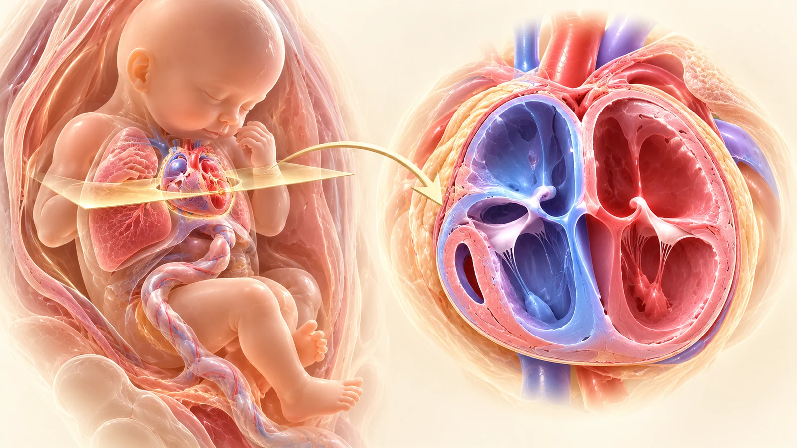
Strict axial alignment across the fetal thorax demonstrating a single, symmetrical lateral rib on each side. The fetal spine is located posteriorly, the sternum anteriorly, and the cardiac apex points toward the left anterior chest wall at 45°. Oblique angulation foreshortens ventricles and artificially induces chamber disproportion.
- Single rib rule: Confirm a single unbroken rib shadow laterally on both sides of the chest
- Avoid oblique planes: Oblique angulation foreshortens ventricles and mimics disproportion or false VSD
- Stomach bubble verification: Confirm gastric bubble is in the left upper quadrant prior to moving probe cephalad into thorax
- Zoom & acoustic focus: Heart should occupy at least 50% of the ultrasound monitor display
Normal Foramen Ovale & Atrial Septal Dynamics
Physiological Right-to-Left Shunt across Septum Primum
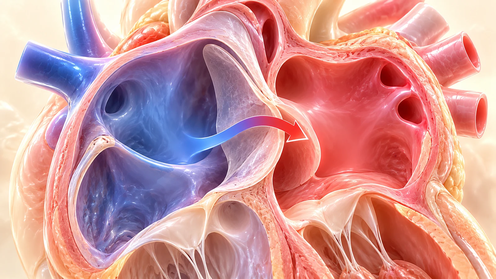
The valve of the fossa ovalis is seen bulging into the left atrium during atrial diastole. In fetal life, approximately one-third of inferior vena cava flow is preferentially shunted across the foramen ovale into the left atrium to supply oxygen-rich blood to the coronary arteries and cerebral circulation.
- Flap excursion: Flap must deflect into the left atrium; excursion should occupy roughly 1/3 to 1/2 of left atrial diameter
- Intact septum primum: Continuous lower rim at crux prevents mistaking normal foramen for a primum ASD
- Redundant aneurysm: Flap touching lateral left atrial wall suggests redundant septum or hypermobility
- Restricted flow warning: Premature restriction/closure presents with right atrial dilation and left-sided hypoplasia
Section 3 — The OBGYNX Abnormal 4CH Diagnostic Fork
Starting Point: "The fetal four-chamber view appears abnormal. What exactly is abnormal?"
The Master Abnormal 4-Chamber Diagnostic Fork
Algorithmic Differential Pathway from First Ultrasound Observation
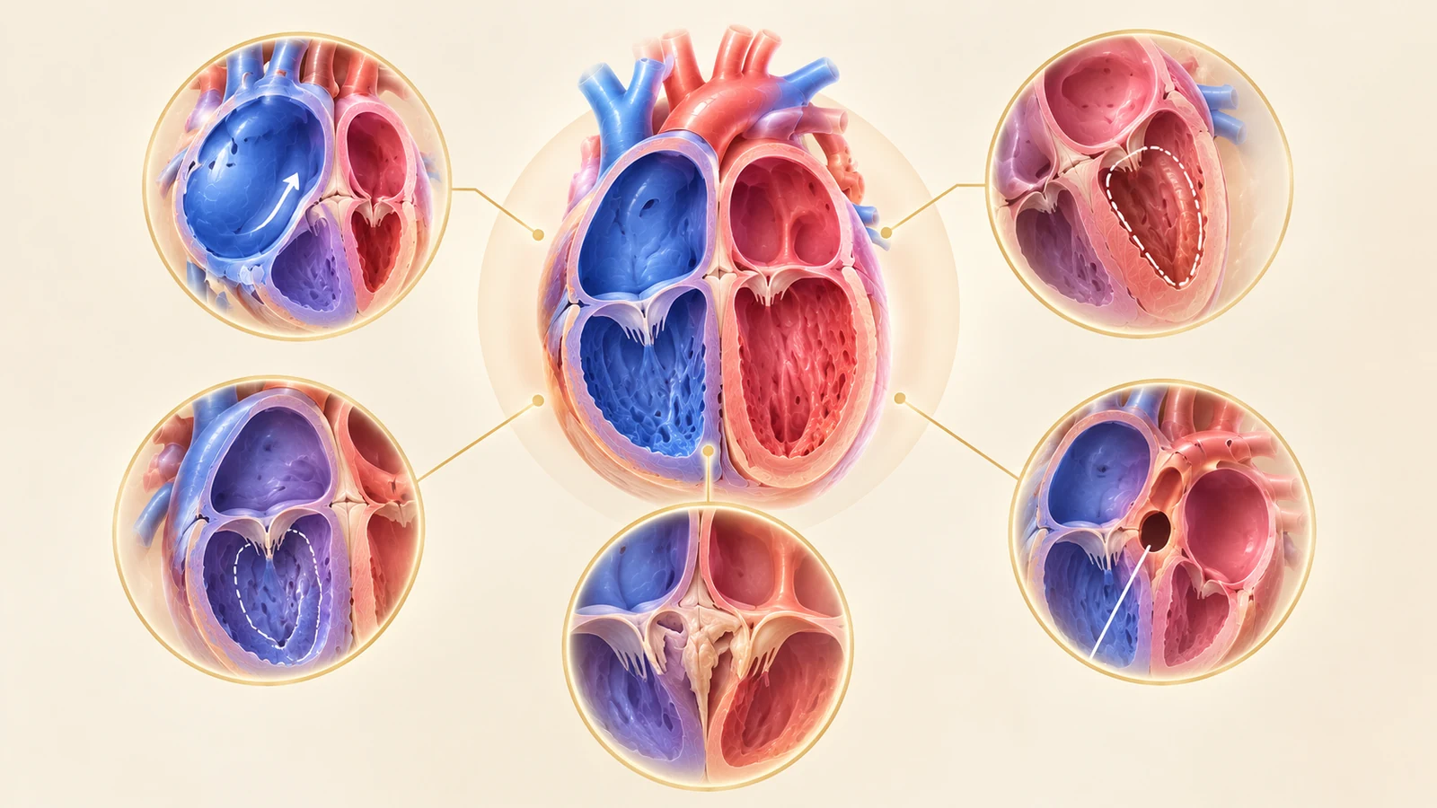
Comprehensive diagnostic bifurcation framework dividing any abnormal four-chamber view into distinct anatomical categories: Chamber Size Disproportion (Small LV vs Small RV), Crux Architecture, Great Vessel Alignment, Septal Defects, Cardiac Axis/Position, and Functional Cardiomegaly.
- Branch A: Cardiac Axis & Position (>65° or <25° deviation)
- Branch B & C: Chamber Disproportion (LV < RV: HLHS, Coarctation, TAPVC)
- Branch D: Small RV (PA-IVS, Tricuspid Atresia, Critical PS)
- Branch E & G: Crux & AV Valve Abnormalities (AVSD, Ebstein Anomaly)
- Branch H & J: Septal Defects & Cardiomegaly / Effusions
Left Ventricle Smaller Than Right (Small LV)
The most frequent and clinically challenging chamber disproportion in fetal cardiology
LV end-diastolic diameter < RV diameter (ratio RV/LV > 1.3 in mid-gestation or > 1.5 in late 3rd trimester). LV cavity narrow or diminutive.
Disproportion with patent mitral and aortic valves; narrow transverse arch and isthmus on 3VT.
Aortic and/or mitral atresia/severe stenosis, globular non-contractile LV, retrograde arch flow.
Thickened, doming aortic valve with poor LV fractional shortening and endocardial fibroelastosis.
Preload redistribution past 34–36 weeks; completely normal outflow tracts and arches.
IUGR, persistent left SVC stealing venous return, or anomalous pulmonary venous return.
- •Mitral valve inflow: normal biphasic laminar flow vs restricted diminutive jet vs absent inflow
- •LV apex formation: does the LV reach the apex, or does the RV form the entire cardiac apex?
- •Aortic root and 3VT: antegrade laminar blue flow (coarctation/normal) vs retrograde red flow (HLHS)
- •Endocardial fibroelastosis: bright, hyperechoic LV endocardial rim indicates high intraventricular pressure
- →LVOT View: evaluate aortic annulus diameter (Z-score) and leaflet mobility
- →3VT View: mandatory to evaluate aortic isthmus caliber and color Doppler directionality
- →Sagittal Aortic Arch View: measure distal transverse arch and isthmus diameters
- ✕Prematurely diagnosing HLHS in an awake patient when the LV is simply small from coarctation
- ✕Reassuring parents that a small LV is 'normal late-term disproportion' without imaging the aortic isthmus
Inadequate left-sided fetal blood volume flow (often secondary to early aortic or mitral inflow limitation or abnormal ductus venosus streaming).
Comprehensive fetal echo; serial growth scans every 2–3 weeks; planned delivery at tertiary pediatric cardiac center with neonatology on standby for PGE1 infusion.
Section 4 — Left Ventricle Is Small: The Disproportion Dilemma
A disproportionately small left ventricle is the single most common reason for emergency fetal cardiology referral. The clinical stakes are immense: misinterpreting severe coarctation as "mild late-term disproportion" can lead to fatal ductal closure at home postnatally, while incorrectly labeling an isolated small LV as Hypoplastic Left Heart Syndrome (HLHS) creates catastrophic parental distress.
1. Classical Hypoplastic Left Heart Syndrome (HLHS): The combination of aortic and/or mitral valve atresia or severe hypoplasia. The LV cavity is slit-like, thick-walled, or completely non-contractile. The cardinal diagnostic sign is retrograde (red) blood flow down the ascending aorta and transverse arch on 3VT imaging, proving the LV cannot generate systemic perfusion pressure.
2. Critical Aortic Coarctation (CoA): The LV is small, but the mitral valve and aortic root are patent with antegrade (blue) forward flow. On the 3VT view, the transverse aortic arch is distinctly narrower than the wide ductal arch. Crucial fact: prenatal detection of coarctation has a high false-positive rate (~20–30%) because late-gestation hemodynamic shifts frequently make the RV appear physiological larger.
3. Critical Aortic Valve Stenosis: The LV is globular and hypertrophied with bright, reflective endocardial fibroelastosis (EFE). Systolic ballooning and turbulent systolic jetting (> 2.5 m/s) with poor contractility are visible. Early identification allows potential in utero fetal aortic valvuloplasty to rescue biventricular circulation!
4. Unbalanced Atrioventricular Septal Defect: The common AV valve is shifted towards the right, directing >60% of inflow to the RV and starving the LV of diastolic volume.
Left Ventricle Smaller Than Right (Ventricular Disproportion)
Visual comparison distinguishing Hypoplastic Left Heart Syndrome (HLHS) with slit-like non-contractile LV and retrograde transverse arch flow from Coarctation of the Aorta (CoA) with patent antegrade valves and isolated aortic isthmus tapering on 3VT view.
Hypoplastic Left Heart Syndrome (HLHS) Spectrum
Small/Slit-Like LV with Aortic/Mitral Atresia
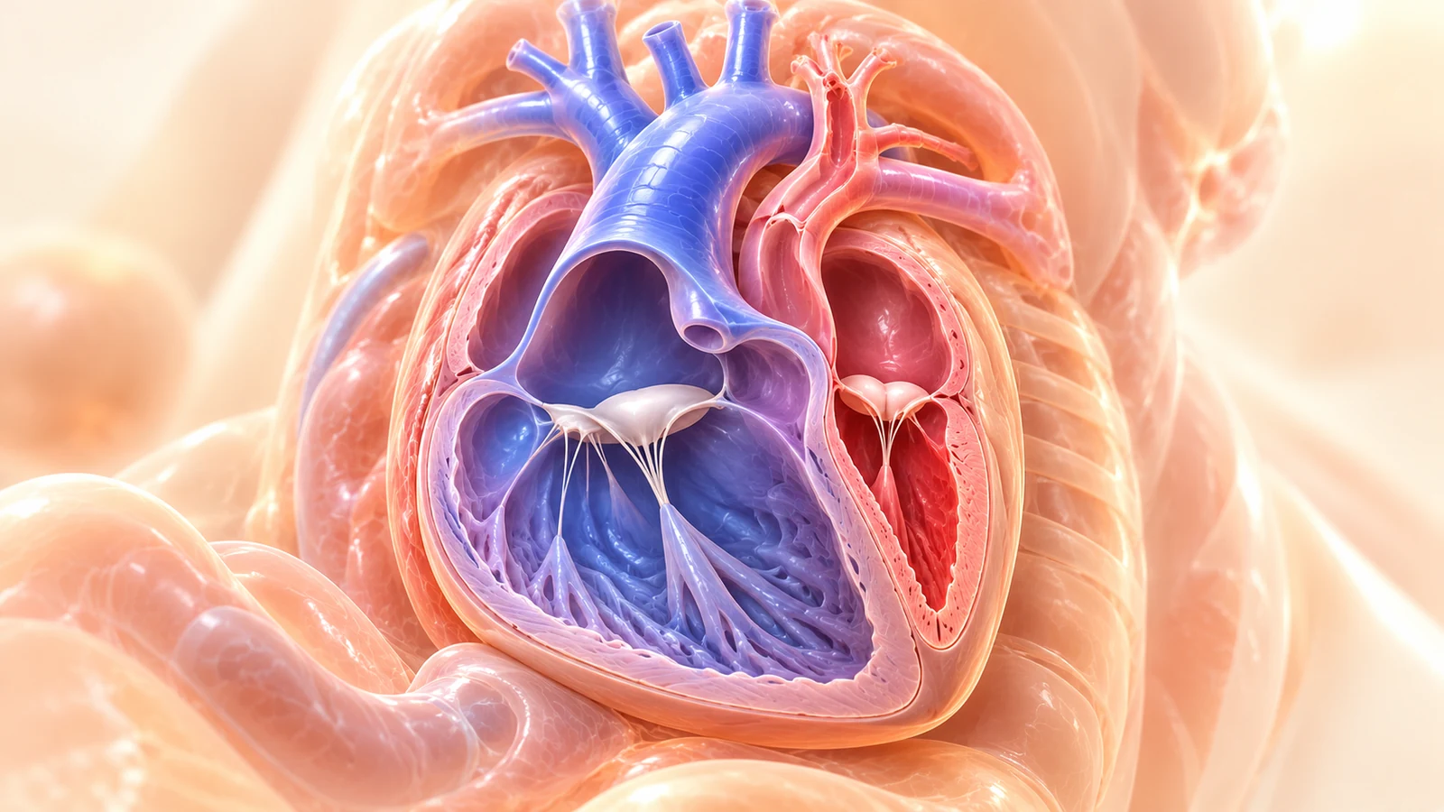
The left ventricle is severely diminutive, globular, or slit-like with thick echogenic endocardial fibroelastosis. The apex is formed entirely by the dilated, hypertrophied right ventricle. Inflow through the mitral valve is absent or severely restricted, and aortic outflow demonstrates retrograde flow on color Doppler.
- Apex formation: Cardiac apex is formed entirely by the right ventricle
- Endocardial fibroelastosis (EFE): Bright echogenic lining of LV wall indicates non-compliant, hypertensive ventricle
- Reversed foramen ovale shunt: Left-to-right interatrial flow if mitral stenosis with intact atrial septum
- Retrograde aortic arch flow: Color Doppler in 3VT demonstrates blue (retrograde) flow down the diminutive transverse arch
Suspected Coarctation of the Aorta (CoA)
Right-Heart Dominance with Patent Mitral Valve & Shelf
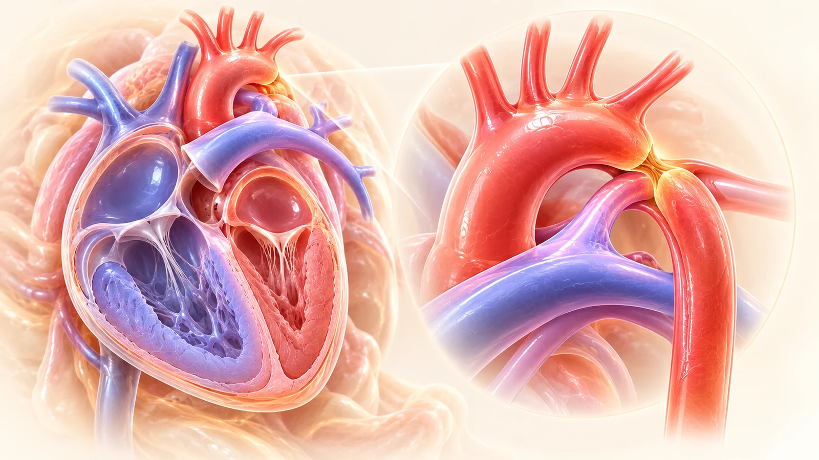
In coarctation of the aorta, the four-chamber view shows ventricular disproportion with a smaller LV and dominant RV. Unlike HLHS, the mitral valve is fully patent with forward biphasic flow across the mitral and aortic valves, but the aortic arch isthmus exhibits a discrete shelf with post-stenotic turbulence.
- Patent mitral valve: Normal biphasic diastolic E/A Doppler wave (distinguishes CoA from mitral atresia in HLHS)
- Late-gestational disproportion: Disproportion frequently becomes pronounced only after 30–32 weeks of gestation
- 3VT view discrepancy: Hypoplastic transverse aortic arch isthmus smaller than ductal arch (isthmus:ductus ratio < 0.6)
- Posterior shelf: High-resolution sagittal sweep demonstrates discrete posterior shelf at aortic isthmus
Section 5 — Right Ventricle Is Small: The Right Hypoplasia Spectrum
When the right ventricle fails to reach the apex and appears noticeably smaller than the left, a primary failure of right heart development or severe inflow/outflow obstruction must be investigated. The major causes include Pulmonary Atresia with Intact Ventricular Septum (PA-IVS), Tricuspid Atresia, and Critical Pulmonary Stenosis.
The RV is heavily hypertrophied with a small or obliterated cavity. Tricuspid valve is diminutive with severe high-velocity regurgitation. The cardinal feature on 3VT is retrograde ductal flow (aorta into pulmonary trunk) supplying the fetal lungs. Look meticulously for ventriculo-coronary sinusoids!
The tricuspid valve orifice is completely absent and replaced by a thick, echogenic fibrous plate. The right ventricle is a small, passive pouch beneath the LV, filled only via a ventricular septal defect. Mandatory single-ventricle surgical management postnatally.
Pulmonary Atresia with Intact Ventricular Septum (PA-IVS)
Small, Concentric Hypertrophic RV with Intact Septum
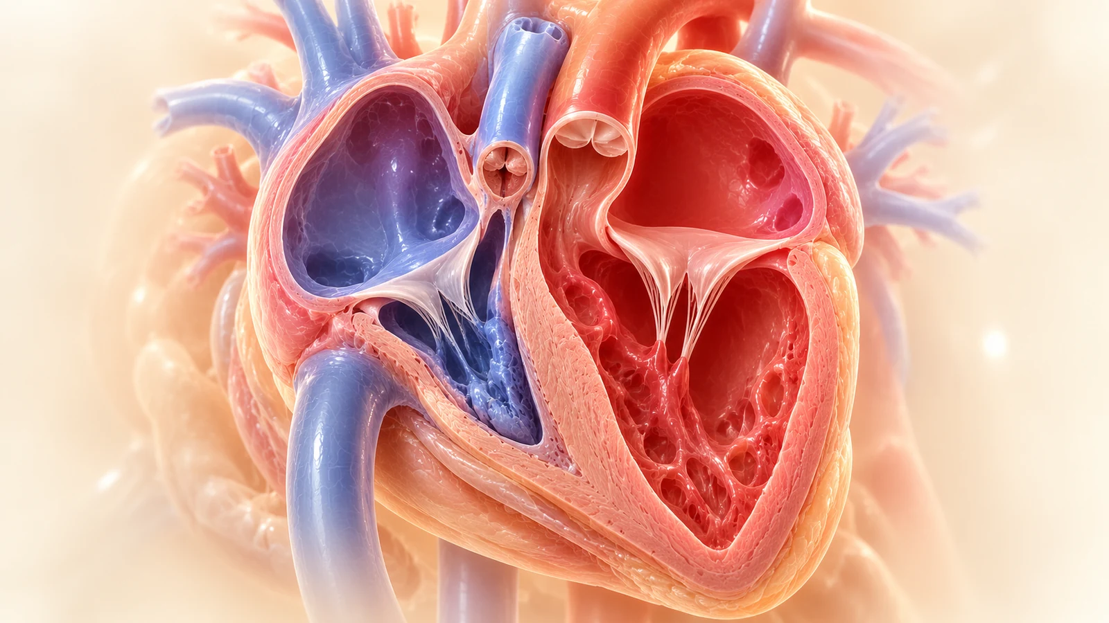
The right ventricle is diminutive with marked concentric myocardial wall hypertrophy, obliteration of the trabecular cavity, and a patent but severely stenotic or dysplastic tricuspid valve. The interventricular septum remains completely intact, and pulmonary outflow is supplied retrogradely via the ductus arteriosus.
- Cavity obliteration: Severe concentric wall thickening with diminutive RV cavity (tripartite vs bipartite)
- Intact ventricular septum: No VSD present (distinguishes PA-IVS from Tetralogy of Fallot with pulmonary atresia)
- High-velocity TR: Doppler displays high-velocity tricuspid regurgitation jet (>3.5–4.5 m/s) due to suprasystemic RV pressure
- Ventriculocoronary arterial connections (sinusoids): Risk of RV-dependent coronary circulation
Tricuspid Atresia: Absent Right Atrioventricular Connection
Imperforate Fibrous Plate with Dominant Single Left Ventricle
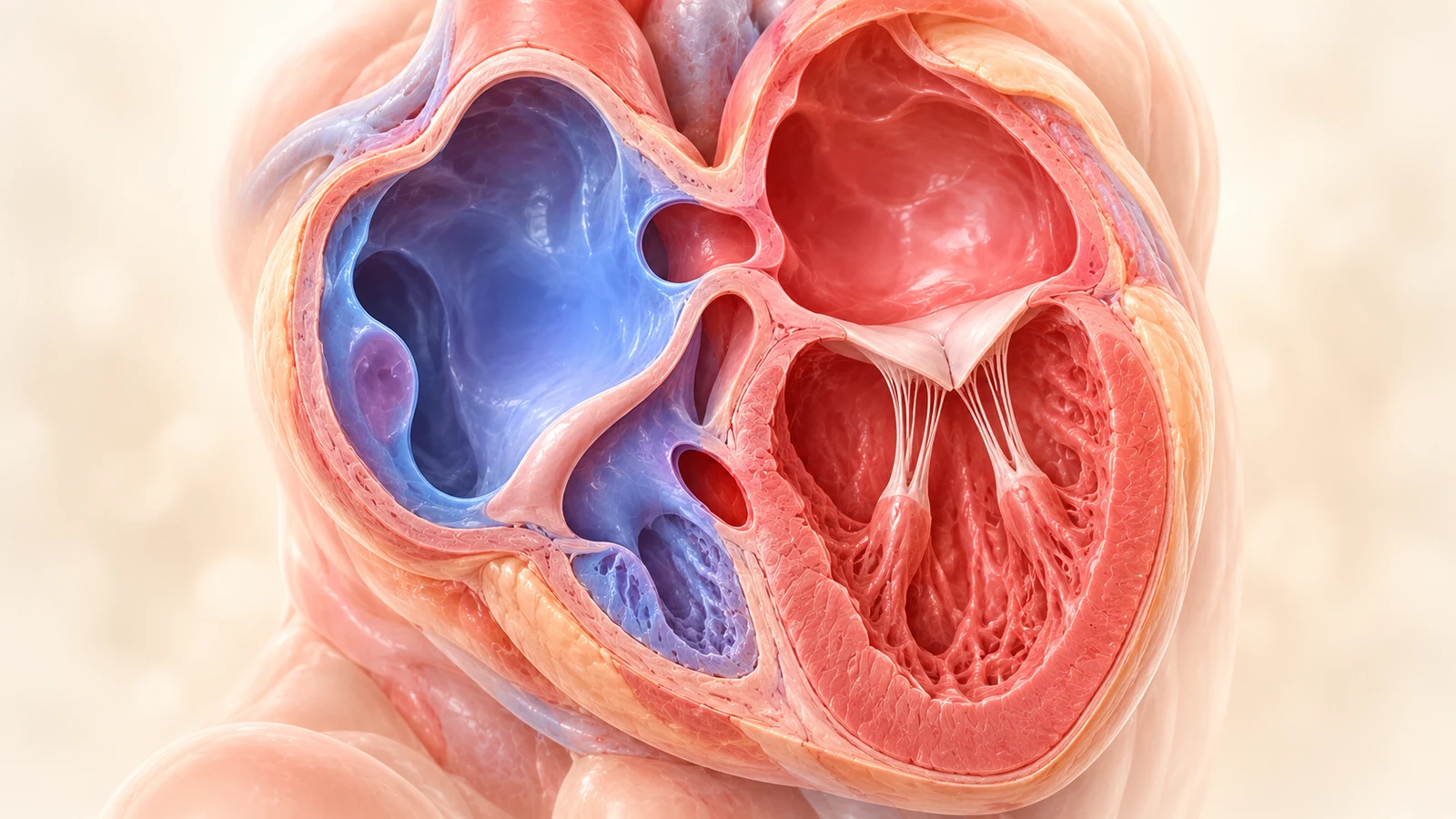
Complete absence of the tricuspid valve orifice, replaced by a dense, hyperechogenic fibrous or muscular floor separating the right atrium from the hypoplastic right ventricle. All systemic venous return from the right atrium must cross an obligatory ASD to reach the dilated left ventricle.
- Imperforate AV floor: Dense fibrous plate replaces tricuspid valve; no color flow into RV
- Obligatory right-to-left atrial shunt: Large ASD or stretched foramen ovale carrying 100% of systemic return
- Ventricular septal defect: Necessary for blood flow to reach hypoplastic RV and pulmonary artery
- Apex dominated by LV: Smooth LV forms cardiac apex entirely; single functioning ventricle physiology
Section 6 — Abnormally Large Right Atrium: Ebstein Anomaly vs Tricuspid Dysplasia
A disproportionately massive right atrium occupying more than half the fetal thorax is an ominous finding. The chief differential is between Ebstein anomaly and isolated tricuspid valve dysplasia, both of which cause torrential tricuspid regurgitation and severe risk of hydrops fetalis.
| Anatomical Feature | Ebstein Anomaly | Tricuspid Valve Dysplasia |
|---|---|---|
| Hinge-Point Offset | Marked apical displacement ≥ 8 mm/m² (> 2 mm) | Normal hinge-point position at the atrioventricular junction |
| Atrialized RV | Present; thin-walled, paradoxical contraction | Absent; entire RV cavity functions as a ventricle |
| Leaflet Morphology | Septal/posterior tethered to wall; anterior redundant "sail-like" | Thickened, nodular, shortened chordae, restricted motion |
| Pulmonary Outflow | Frequent "functional" pulmonary atresia from low forward pressure | Forward flow usually preserved unless RV pressure is exhausted |
Ebstein Anomaly of the Tricuspid Valve
Failure of Delamination & Downward Leaflet Displacement
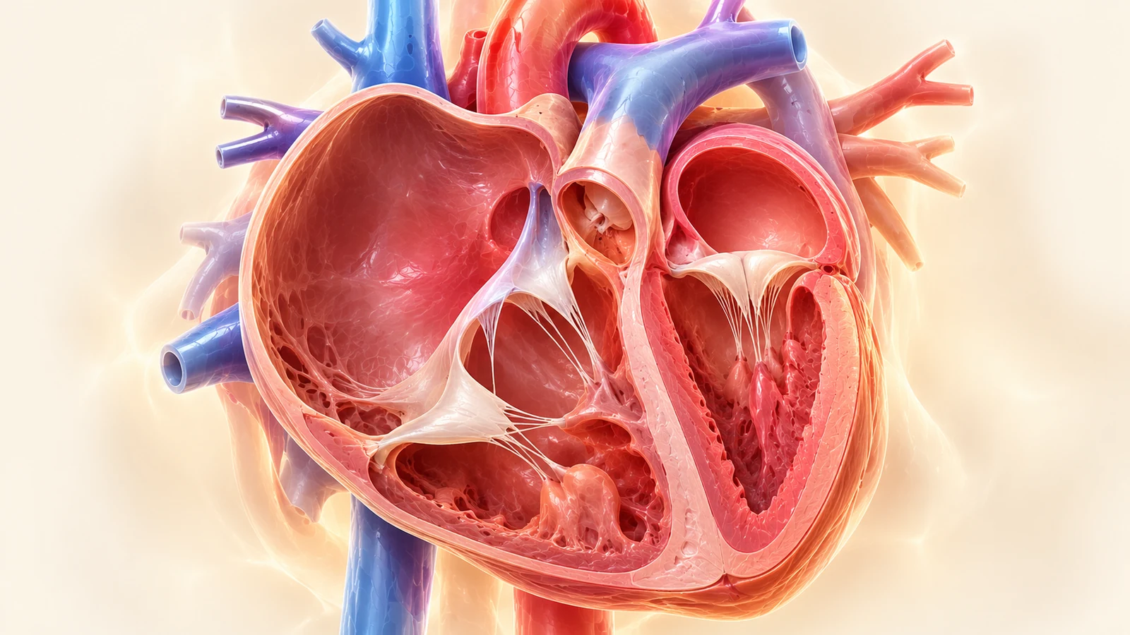
Marked apical displacement of the septal and posterior tricuspid leaflets into the right ventricular cavity (>8 mm/m² BSA or >1.5–2.0 mm). This creates a massive 'atrialized' right ventricle, a small functional right ventricle, and a giant right atrium with torrential regurgitation.
- Apical displacement: Septal leaflet hinged significantly lower on septum than anterior mitral leaflet
- Atrialized RV: Thin, dyskinetic ventricular wall between true annulus and displaced valve hinge point
- Torrential tricuspid regurgitation: Wide, pansystolic regurgitant jet originating deep within the ventricular cavity
- Functional pulmonary atresia: Severe TR prevents forward opening of pulmonary valve in severe cases
Tricuspid Valve Dysplasia
Thickened, Nodular Leaflets Hinged at Normal Annulus
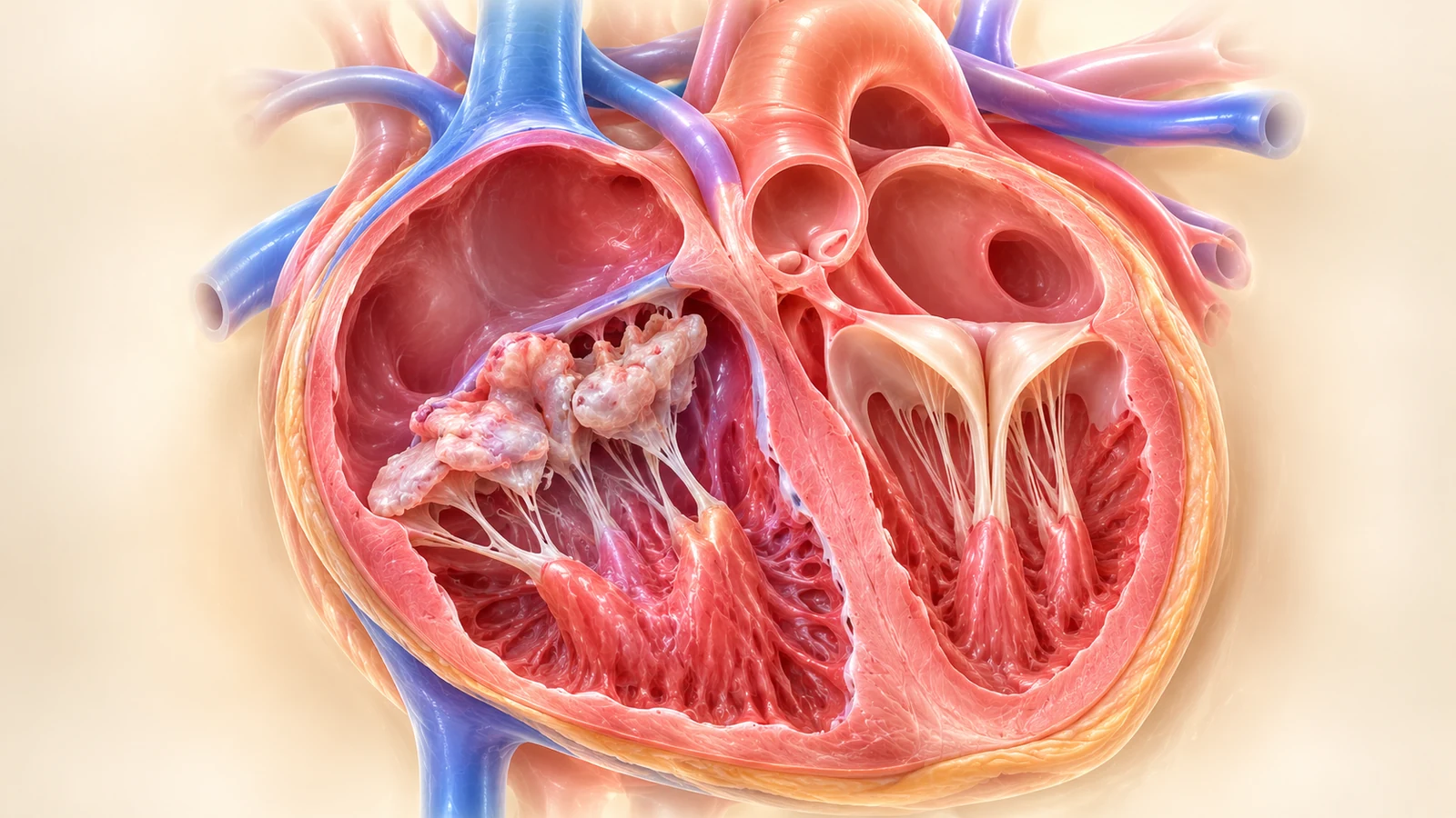
Unlike Ebstein anomaly, the tricuspid valve leaflets in tricuspid dysplasia insert at their normal anatomical annulus without apical downward displacement. The leaflets are thickened, retracted, and nodular with restricted mobility, producing severe tricuspid regurgitation and massive right atrial dilatation.
- Normal annular insertion: No apical displacement of septal leaflet; crux offset is preserved
- Leaflet morphology: Thickened, irregular, echogenic leaflets with restricted coaptation
- Massive right atrial dilation: Right atrium expands to occupy >50% of thoracic cross-section
- Differentiation from Ebstein: Lack of atrialized RV chamber confirms isolated dysplasia
Section 7 — Abnormal AV Valves and Cardiac Crux: The AVSD Spectrum
The cardiac crux is the anatomical junction where the atrial septum, ventricular septum, and atrioventricular valves meet. In normal anatomy, the tricuspid valve inserts 1–2 mm more apical than the mitral valve. Complete loss of this offset—where both valve components insert in a straight linear plane or form a single common orifice—is pathognomonic for an atrioventricular septal defect.
Single common AV valve with 5 leaflets spanning across both ventricles. Large primum ASD above and inlet VSD below. Strongly associated with Trisomy 21 (~60%).
Two separate valve orifices but linear insertion at crux with cleft anterior mitral leaflet and primum ASD. Ventricular septum is intact.
Common AV valve commits predominantly to one ventricle, causing severe contralateral hypoplasia. Requires single-ventricle palliation postnatally.
Complete Atrioventricular Septal Defect (cAVSD)
Loss of Crux Offset with Single Common AV Valve
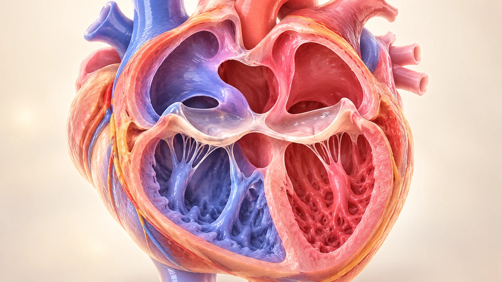
The normal cardiac crux is absent. A single common five-leaflet AV valve spans across both ventricular inlets. In diastole, a large primum atrial septal defect and unrestrictive inlet ventricular septal defect are visualized forming an iconic 'bare area' in the center of the heart.
- Linear insertion: Both AV valve components insert at exactly the same level across the ventricular crest
- Common valve orifice: Open valve reveals single shared orifice in diastole; cleft left AV valve
- Primum ASD + Inlet VSD: Central H-shaped or cross-shaped deficit at the junction of septa
- Trisomy 21 association: Up to 60% of fetuses with complete AVSD have Down syndrome
Section 8 — Suspected Septal Defects: True Defect vs Ultrasound Artifact
Evaluating the ventricular and atrial septa requires understanding acoustic physics. The thin membranous septum readily causes acoustic drop-out in apical views, creating false VSD alarms. Conversely, small muscular VSDs may only be detectable on perpendicular high-resolution color Doppler sweeps.
A genuine ventricular septal defect displays hyperreflective, rounded, blunted margins at the edge of the muscular septum—known as the T-sign. In contrast, acoustic drop-out artifact displays jagged, fading, non-reflective borders that vanish completely when the probe is rotated 90° into a subcostal plane perpendicular to the septum!
Ventricular Septal Defect (VSD) vs Acoustic Drop-Out
Perpendicular Interrogation & The Diagnostic 'T-Sign'
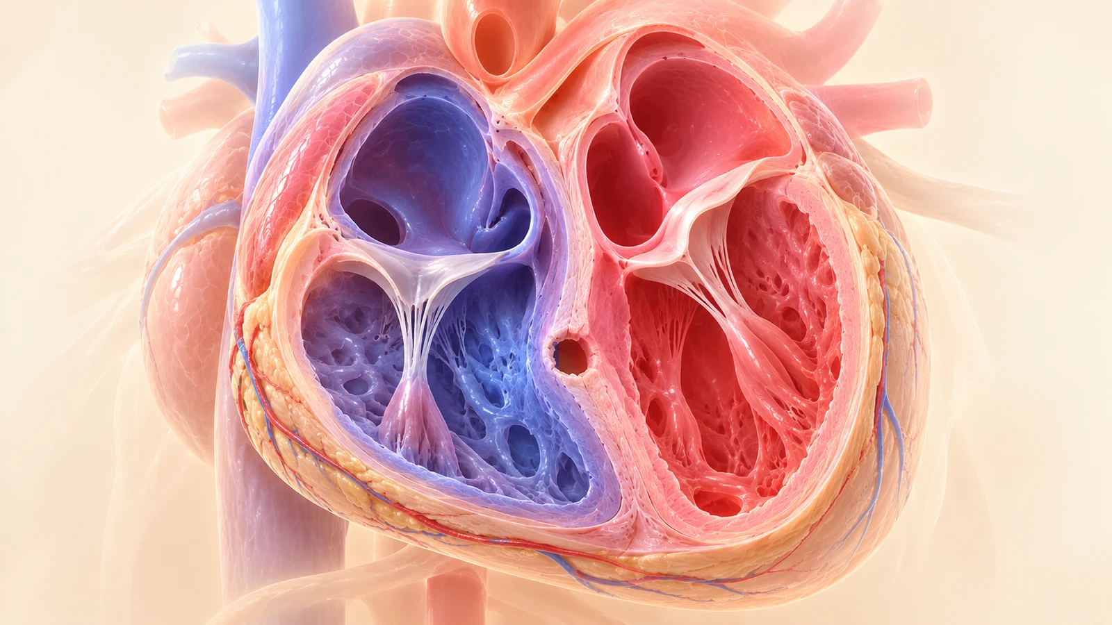
True ventricular septal defect demonstrated with an ultrasound beam oriented perpendicular to the septum. Bright echogenic capping ('T-sign') borders the defect margins on gray-scale, and bidirectional or left-to-right color Doppler flow confirms anatomical discontinuity.
- Beam perpendicularity: Never diagnose a VSD from an apical view alone due to acoustic drop-out
- The 'T-Sign': Distinct thickening and echogenic border on both sides of the septal gap confirms true anatomical margins
- Color Doppler persistence: Adjust color scale (PRF ~40–50 cm/s) to visualize jet crossing septum
- Inlet vs perimembranous vs muscular: Correlate with 4CV, LVOT, and short-axis views to localize precise defect subtype
The foramen ovale is an obligatory physiological fetal communication shunting oxygenated blood from the placenta to the left heart. Never label a normal foramen ovale an "atrial septal defect (ASD)." True primum ASDs occur at the crux as part of AVSD; true sinus venosus and secundum ASDs are generally impossible to diagnose with certainty prenatally because the flap is naturally patent.
Section 9 — Abnormal Cardiac Axis, Situs and Position
The normal cardiac axis points into the left hemithorax at 45° ± 20° (ranging 25° to 65°). An axis outside this range is one of the most sensitive screening markers for congenital heart disease, even when four-chamber anatomy appears deceptively normal.
Cardiac apex points to the right hemithorax. If accompanied by situs inversus totalis, CHD risk is ~5–10%. If situs is solitus (isolated dextrocardia), CHD risk exceeds 90%!
The heart is displaced into the right chest by a space-occupying lesion (CDH, CPAM, massive left pleural effusion), but intrinsic axis may still remain oriented leftward.
Axis rotated hyper-leftward. High-yield red flag for conotruncal malformations (Tetralogy of Fallot, ccTGA) or coarctation of the aorta.
Fetal Cardiac Axis Measurement & Deviation
Spine-Sternum Line Reference (Normal 45° ± 20°)
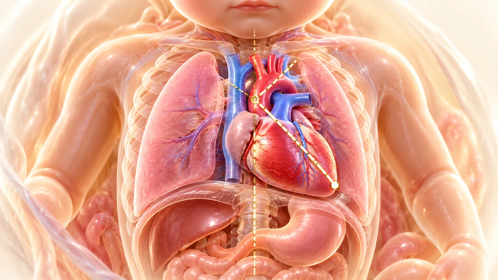
Demonstration of cardiac axis measurement in a true axial thoracic slice. A reference line is drawn from the center of the vertebral body to the anterior midline sternum. A second line is traced along the interventricular septum. Normal axis ranges between 25° and 65°.
- Measurement protocol: Draw mid-sagittal reference line through spine and anterior sternum
- Normal angle: 45° with normal standard deviation of ± 20° (25°–65°)
- Extreme levo-axis (>65°): Strongest indicator of conotruncal CHD (TOF, truncus arteriosus, DORV, coarctation)
- Dextro-axis (<25° or pointing right): Suggests dextrocardia, congenital diaphragmatic hernia, or heterotaxy
Thoraco-Abdominal Situs & Lateral Orientation
Excluding Heterotaxy, Isomerism, and Dextroposition
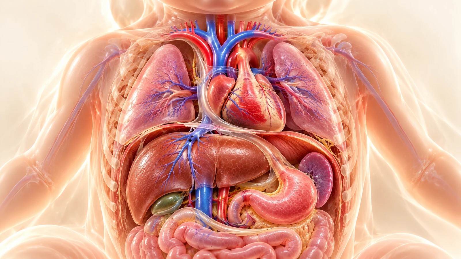
Systematic assessment of abdominal and thoracic visceral situs. In situs solitus, the stomach bubble, cardiac apex, descending aorta, and spleen reside on the left, while the liver, IVC, and gallbladder reside on the right.
- Fetal lie verification: Always determine head position and spinal rotation before declaring left vs right
- Gastric-cardiac concordance: Gastric bubble on left + cardiac apex on left = situs solitus
- Discordance warning: Stomach right + apex left (or vice versa) raises suspicion for heterotaxy / isomerism
- Extracardiac displacement: Dextroposition with normal leftward axis implies external push (CDH, CCAM) or pull (pulmonary hypoplasia)
Section 10 — Cardiomegaly and Abnormal Cardiac Function
Not all cardiomegaly is structural congenital heart disease! When the cardiothoracic ratio (circumference > 0.50 or area ratio > 0.35) is elevated, the fetal medicine specialist must investigate high-output volume overload, arrhythmias, and cardiomyopathies.
- Fetal Anemia: Rh alloimmunization or Parvovirus B19 (check MCA-PSV > 1.5 MoM).
- Arteriovenous Shunts: Sacrococcygeal teratoma, vein of Galen malformation, or placental chorioangioma.
- Twin-to-Twin Transfusion: Recipient twin developing hypervolemic hypertrophy and hydrops.
- Sustained Tachyarrhythmias: Atrial flutter or SVT > 220 bpm causing rate-related cardiomyopathy.
- Complete Atrioventricular Heart Block: Maternal anti-Ro/SSA antibodies causing autoimmune myocarditis.
- Cardiac Tumors: Rhabdomyomas protruding into chambers (associated with Tuberous Sclerosis).
Cardiomegaly: Cardio-Thoracic Circumference & Area
Distinguishing Structural CHD from High-Output Heart Failure
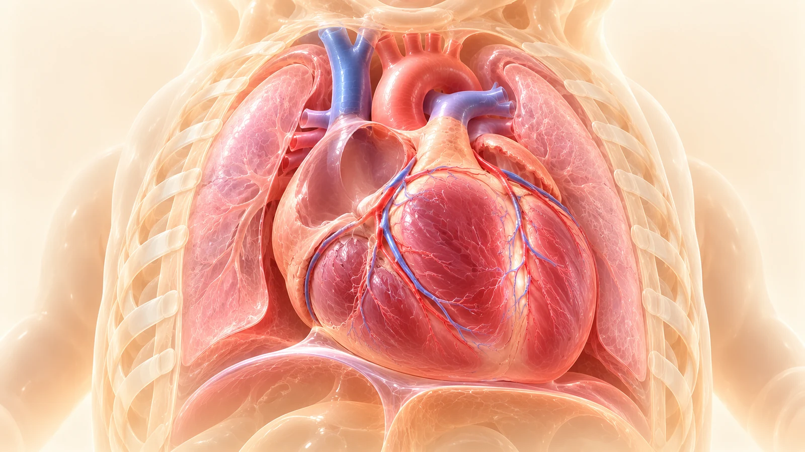
Cardiomegaly confirmed on axial 4CV. The heart occupies more than one-third of the thoracic area or exceeds a cardio-thoracic circumference ratio of 0.50. The lungs are compressed laterally and the cardiac apex pushes firmly against the left anterior ribs.
- Cardiothoracic circumference ratio (CTR): Circumference of heart / Circumference of thorax > 0.50 is abnormal
- Cardiothoracic area ratio: Normal is ~0.33 (one-third); >0.40 indicates definite cardiomegaly
- Etiology breakdown: Severe valve regurgitation (Ebstein), arteriovenous fistulas (Vein of Galen), fetal anemia, TTTS, or maternal diabetes
- Pulmonary hypoplasia risk: Chronic thoracic crowding by an enlarged heart impairs fetal lung alveolar development
Pericardial Fluid: Physiological Halo vs Pathologic Effusion
Quantifying Rim Thickness & Evaluating Myocardial Squeezing
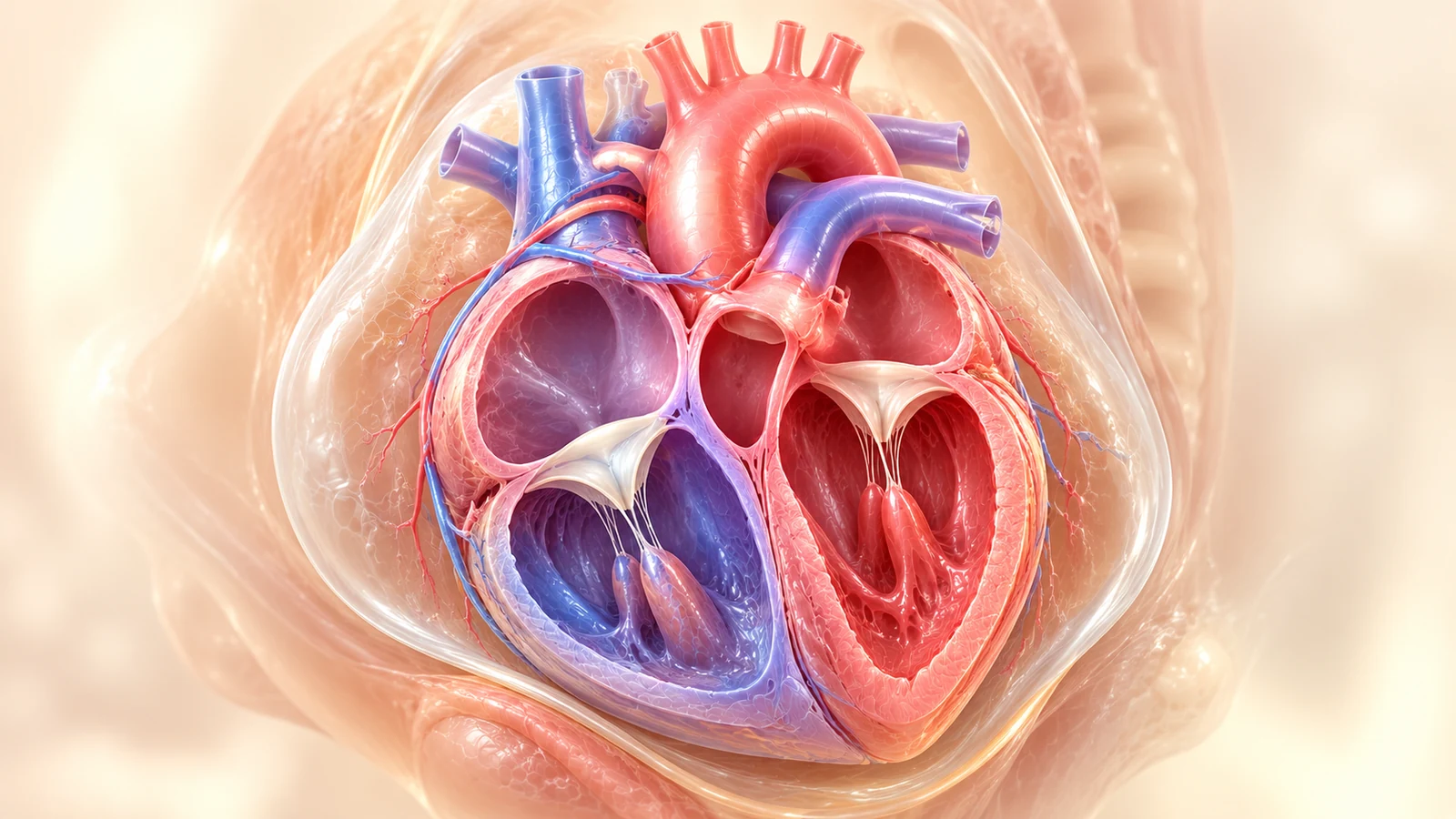
Assessment of pericardial space. A small anterior fluid stripe (<2 mm) localized to the atrioventricular groove is normal. In contrast, circumferential fluid extending behind the left atrium and around the apex measuring >2 mm indicates pathologic pericardial effusion.
- Physiological threshold: Trace fluid stripe < 2 mm confined to anterior AV groove is benign
- Pathologic effusion: Circumferential fluid > 2 mm extending behind posterior myocardial wall
- Cardiac tamponade signs: Diastolic right atrial collapse, ventricular filling impairment, and pulsatile umbilical venous flow
- Hydrops survey: Look for accompanying pleural effusion, ascites, and skin edema (>5 mm)
Section 11 — Abnormal Venous Return: Dilated CS, TAPVC & PLSVC
Systemic and pulmonary venous anomalies frequently hide behind a normal-appearing four-chamber view. However, subtle clues in the atrioventricular groove and the 3VT view allow early detection.
Occurs in ~0.5% of the general population and up to 5% of fetuses with CHD. On the 4CV, it produces a dilated coronary sinus visible as an anechoic oval structure in the posterior AV groove. On the 3VT view, it manifests as a 4th vessel located to the extreme left of the pulmonary artery.
In TAPVC, all pulmonary veins fail to connect to the left atrium, draining instead via a common confluence to the innominate vein (supracardiac), coronary sinus (cardiac), or portal system (infracardiac). Warning: The four-chamber view can appear normal or show only subtle left atrial hypoplasia. You MUST document individual pulmonary veins entering the posterior LA wall!
Persistent Left Superior Vena Cava (PLSVC)
Dilated Coronary Sinus in Posterior AV Groove & 4 Vessels on 3VT
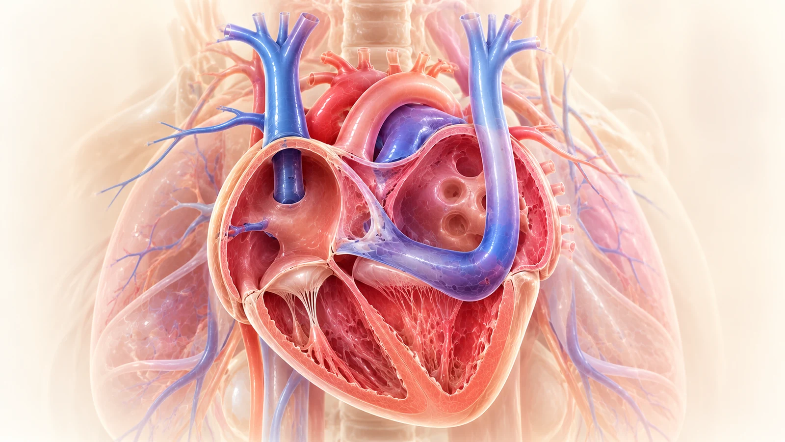
In the four-chamber view, a dilated, round or tubular hypoechoic structure is visualized in the posterior left atrioventricular groove. On the three vessels and trachea (3VT) view, a fourth vessel is seen situated to the left of the pulmonary artery.
- Posterior AV groove: Dilated coronary sinus (>2–3 mm) mimics an atrial septal defect or anomalous pulmonary vein
- 3VT view confirmation: 4 vessels observed: Left SVC, Pulmonary Artery, Aorta, Right SVC (from left to right)
- Association with coarctation: PLSVC is found in ~10–15% of fetuses with coarctation of the aorta
- Isolated benign variant: In the absence of other CHD or aneuploidy, isolated PLSVC is an asymptomatic vascular variant
Total Anomalous Pulmonary Venous Connection (TAPVC)
Absence of Left Atrial Venous Incorporation & Retrocardiac Confluence
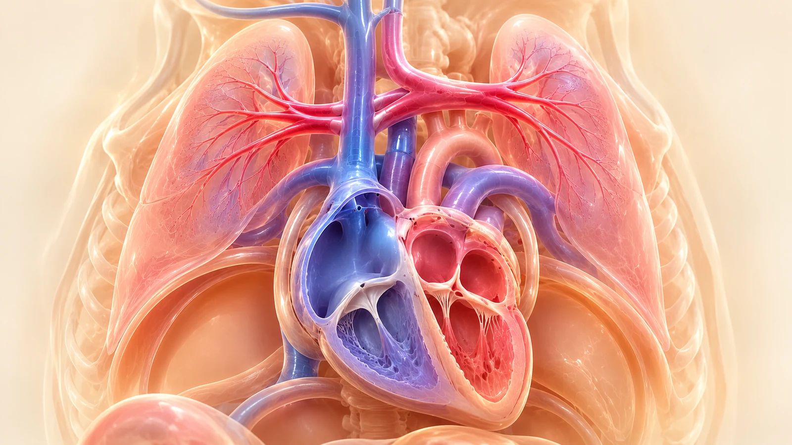
In TAPVC, the posterior wall of the left atrium is entirely smooth without venous dimples. A dilated retrocardiac venous confluence ('collector') is visualized between the left atrium and descending aorta, draining via a vertical vein into the systemic circulation.
- Smooth posterior LA wall: Failure to demonstrate at least two pulmonary veins entering left atrium on color Doppler
- Retrocardiac collector: Echo-free confluence pouch positioned behind left atrium anterior to descending aorta
- Increased LA-aorta distance: Increased space between posterior left atrial wall and descending thoracic aorta
- Pulsatile venous waveform: Spectral Doppler shows continuous, high-velocity or turbulent vertical vein flow
Section 12 — The Normal 4CH Trap: Major CHD with Pristine 4CH Anatomy
A normal four-chamber view is comforting, but it can be a perilous false reassurance. An estimated 40–50% of critical congenital heart defects that require immediate surgical intervention or catheterization in the first days of life present with a completely normal 4CV!
| Congenital Defect | Why 4CV Looks Normal | Diagnostic View That Catches It | Key Ultrasound Finding |
|---|---|---|---|
| D-Transposition (d-TGA) | Chambers and crux are completely symmetrical | LVOT & RVOT Views | Parallel great arteries; aorta anterior from RV; lack of crossover |
| Tetralogy of Fallot (TOF) | Small membranous/outlet VSD often missed on 4CV | LVOT & 3VT Views | Overriding aorta (>50% over IVS); diminutive pulmonary trunk |
| Truncus Arteriosus | Ventricles equal size; VSD obscured on apical 4CV | LVOT & 3VV Views | Single truncal vessel overriding septum; pulmonary arteries branch directly from trunk |
| Coarctation of Aorta | Chamber disproportion may be absent in mid-trimester | 3VT & Sagittal Arch | Narrow transverse arch/isthmus; shelf in descending aorta |
| Vascular Rings (RAA / DAA) | Intracardiac anatomy is 100% completely normal | 3VT View | Right-sided aortic arch; 'U-sign' encircling the trachea |
d-Transposition of the Great Arteries (d-TGA)
Pristine Four-Chamber View Concealing Parallel Outflow Tracts
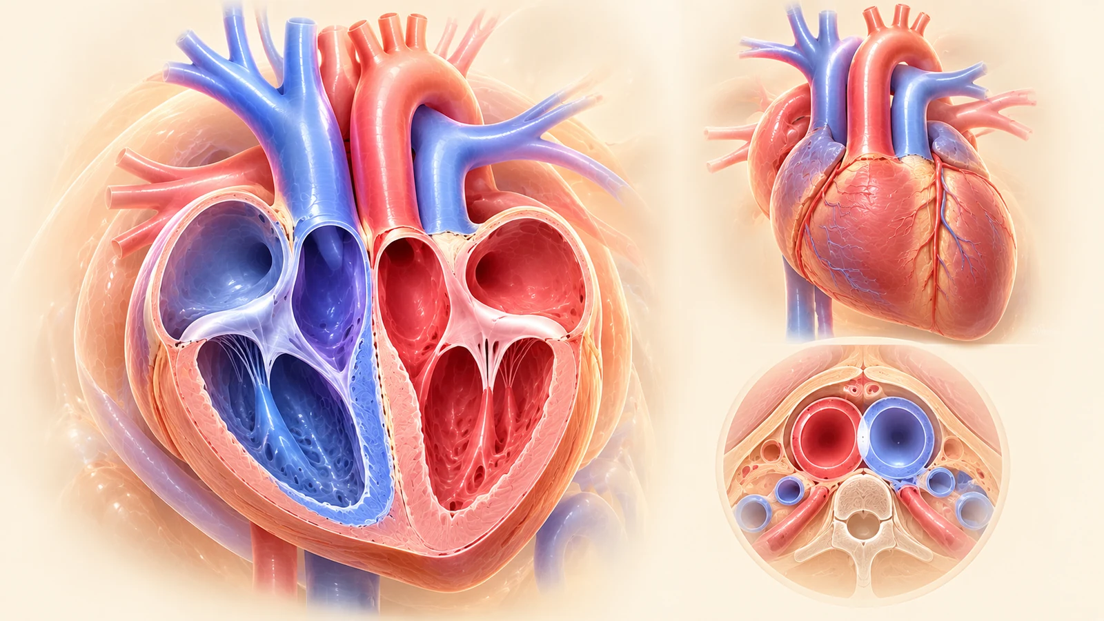
The classic 'Normal 4-Chamber Trap'. The four-chamber view is anatomically flawless with normal chamber sizes and crux. However, angling cephalad into the outflow tracts reveals the aorta arising aberrantly from the right ventricle and the pulmonary trunk arising from the left ventricle, running parallel without normal crossover.
- Normal 4CV: Symmetrical chambers and intact crux provide false reassurance in >95% of screening scans
- Loss of crossover: Normal outflow tracts cross each other at 90°; in d-TGA they run in parallel 'gun-barrel' fashion
- Aorta anterior & right: Bifurcation of posterior pulmonary artery confirms origin from left ventricle
- Emergency postnatal needs: Balloon atrial septostomy (Rashkind) and arterial switch operation (Jatene) in first weeks of life
Tetralogy of Fallot (TOF) & Conotruncal Malformations
Subtle or Normal 4CV with Overriding Aorta on Outflow Views
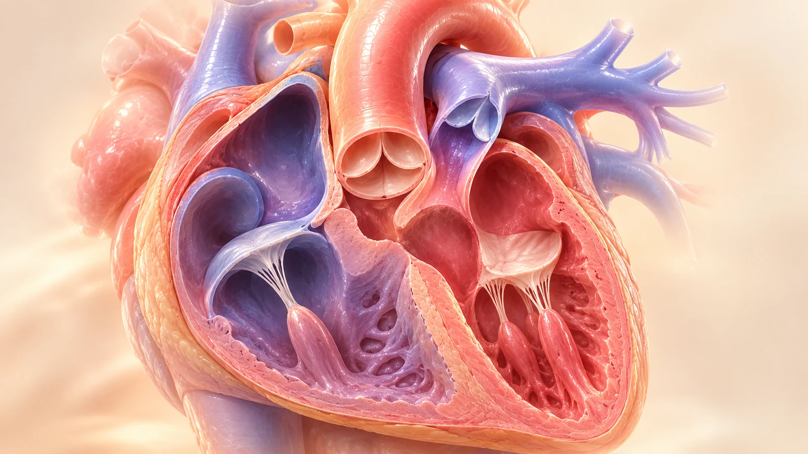
The four-chamber view in mid-gestation may appear normal, or show mild levo-axis deviation. Angling into the LVOT view exposes a large anterior malalignment VSD with an enlarged, overriding aorta straddling the ventricular crest, accompanied by RVOT hypoplasia on the 3VV view.
- 4CV appearance: Often unremarkable in early/mid second trimester; right ventricular hypertrophy is absent prenatally
- Cardiac axis marker: Axis deviation (>65°) is often the only initial clue on the 4CV
- Overriding root: LVOT view clearly shows aortic root overriding ventricular septum by 25–50%
- 3VV view discrepancy: Pulmonary trunk is hypoplastic compared to the dilated ascending aorta
Section 13 — Master Differential Diagnosis Table
A comprehensive clinical desk-reference mapping every abnormal four-chamber ultrasound finding directly to its prioritized differentials, defining signs, essential next views, and common diagnostic traps:
| Abnormal 4CH Finding | Possible Diagnoses | Key Distinguishing Features | Essential Additional Views | Important Pitfalls |
|---|---|---|---|---|
| Small Left Ventricle (RV > LV) | Coarctation of aorta, HLHS, critical AS, late-gestation disproportion | Patent valves & antegrade 3VT flow (CoA) vs atretic valves & retrograde 3VT flow (HLHS) | 3VT, Sagittal Arch, LVOT | Calling HLHS when aorta is patent; missing late coarctation |
| Small Right Ventricle (LV > RV) | PA-IVS, Tricuspid atresia, critical PS, unbalanced AVSD | Thick hypertrophic RV & retrograde ductus (PA-IVS) vs imperforate TV membrane (TA) | RVOT, 3VT, Short-Axis | Missing ventriculocoronary sinusoids in PA-IVS |
| Giant Right Atrium | Ebstein anomaly, tricuspid dysplasia, severe functional TR | Apical TV displacement > 2 mm & atrialized RV (Ebstein) vs normal hinge point (dysplasia) | RVOT, Ductal Arch, M-Mode | Functional pulmonary atresia mistaken for structural atresia |
| Linear Crux (Loss of Offset) | Complete AVSD, partial AVSD, unbalanced AVSD | Common AV valve spanning primum ASD & inlet VSD; cleft mitral leaflet | LVOT ('goose-neck'), Plane 1 | Mistaking for isolated VSD; missing Trisomy 21 association |
| Septal Drop-Out | Acoustic artifact (pseudo-VSD) vs true perimembranous / muscular VSD | Persists in perpendicular subcostal view with 'T-sign' margins and color Doppler shunt | Subcostal 4CV, LVOT | Diagnosing VSD on apical 4CV alone without perpendicular confirmation |
| Abnormal Axis (>65° or <25°) | Conotruncal anomalies (TOF, TGA), heterotaxy, CDH/CPAM displacement | Overriding aorta / parallel vessels vs intrathoracic herniated viscera | Plane 1 (Situs), LVOT, RVOT, 3VT | Dismissing abnormal axis as 'normal variation' |
| Cardiomegaly (CTR > 0.50) | Fetal anemia, AV shunts, SVT/heart block, cardiomyopathy, hydrops | Elevated MCA-PSV > 1.5 MoM (anemia) vs heart rate > 220 or < 60 bpm | MCA Doppler, Ductus Venosus, M-Mode | Assuming all cardiomegaly is structural cardiac disease |
Section 14 — 12 Clinical Diagnostic Pearls
A small LV does not equal HLHS. Always check the 3VT view: if transverse arch flow is antegrade (blue), the LV is functional and coarctation is the primary suspect. If retrograde (red), HLHS is confirmed.
Never diagnose a VSD with the ultrasound beam pointing straight at the apex. Turn the transducer 90° into a subcostal plane to look for true myocardial 'T-sign' margins.
A cardiac axis > 65° in the presence of a completely normal 4CV carries a 30–40% risk of underlying conotruncal or arch anomaly.
Linear insertion of atrioventricular valves with a primum ASD carries an ~60% probability of Down syndrome. Amniocentesis with chromosomal microarray is strongly indicated.
D-TGA, Tetralogy of Fallot, and Truncus Arteriosus almost never alter the 4CV. An obstetric screening exam that stops at the 4CV misses nearly half of surgical CHD!
Ebstein anomaly with severe TR and high Celermajer index (>1.0) carries a >80% risk of developing fetal hydrops and intrauterine demise without maternal-fetal medical therapy.
A cystic structure in the posterior left AV groove is almost always a dilated coronary sinus secondary to a Persistent Left SVC. Confirm by finding 4 vessels in the 3VT view.
In severe Ebstein anomaly, the pulmonary valve may appear atretic simply because severe TR dissipates RV pressure. Distinguish functional atresia by checking for structurally normal valve leaflets.
After 34 weeks, an RV/LV ratio up to 1.35 is physiologically normal. Do not trigger emergency deliveries for mild late disproportion if outflow tracts and arch flows are pristine.
Always obtain an MCA peak systolic velocity in fetuses with unexplained cardiomegaly to rule out severe fetal anemia before assuming intrinsic cardiomyopathy.
A brightly hyperechoic, glittering left ventricular endocardial lining indicates chronic severe intraventricular hypertension, seen in critical aortic stenosis and evolving HLHS.
Never counsel parents on prognosis or surgical mortality based on an abnormal 4CV alone. A complete segmental fetal echocardiogram by a pediatric cardiologist is mandatory.
Section 15 — The 12-Point Step-by-Step Diagnostic Protocol
- Step 1: Confirm Laterality & Situs: Upper abdominal view (Plane 1) verifying stomach and heart apex concordantly on the left.
- Step 2: Verify True Axial Thoracic Cut: Symmetrical single ribs on each lateral wall without oblique foreshortening.
- Step 3: Measure Cardiac Size & Axis: CTR < 0.50; axis 45° ± 20° into left chest; 2/3 of heart in left hemithorax.
- Step 4: Identify Morphological RV vs LV: Coarse trabeculae & moderator band = RV; smooth conical apex = LV.
- Step 5: Compare Ventricular Sizing (1:1 Ratio): Rule out chamber disproportion (RV/LV ratio normally 1.0–1.2).
- Step 6: Evaluate Cardiac Crux & AV Valve Offset: Tricuspid septal leaflet inserts 1–2 mm more apical than mitral valve.
- Step 7: Interrogate Interventricular Septum: Examine in perpendicular subcostal plane with color Doppler to exclude VSD.
- Step 8: Confirm Atrial Septum & Foramen Ovale Flap: Flap moves exclusively into the left atrium; primum septum intact.
- Step 9: Document Pulmonary Venous Insertion: At least two pulmonary veins verified entering posterior left atrium.
- Step 10: Assess Myocardial Squeezing & Rhythm: Symmetrical ventricular contraction; regular 1:1 AV conduction (110–160 bpm).
- Step 11: Mandatory Outflow & 3VT Sweeps: Sweep cephalad to confirm LVOT septoaortic continuity, RVOT crossover, and 3VT left-sided V-sign.
- Step 12: Formulate Structured Referral: Document precise morphological finding and arrange pediatric cardiology fetal echo.
Section 16 — Clinical Reasoning Quiz: 8 Critical 4-Chamber Diagnostic Scenarios
Board-level interactive clinical cases evaluating chamber balance, crux architecture, septal interrogation, and conotruncal traps.
Which diagnostic interpretation and management pathway is clinically correct?
The Abnormal Four-Chamber View: From Observation to Differential Diagnosis
Prenatal fetal cardiac diagnosis is a discipline of sequential deduction. When confronted with an abnormal four-chamber view, follow the OBGYNX Five-Stage Diagnostic Method:
Primary Scientific References & Literature Sources
- Carvalho JS, et al. ISUOG Practice Guidelines (updated): fetal cardiac screening. Ultrasound Obstet Gynecol 2023; 61: 788–803. [DOI: 10.1002/uog.26224]
- American Society of Echocardiography. Guidelines and Recommendations for Performance of the Fetal Echocardiogram. J Am Soc Echocardiogr 2023. [ASE Guidelines 2023]
- ISUOG VISUOG: Abnormal Four-Chamber View. [ISUOG VISUOG Reference]
- Donofrio MT, et al. Diagnosis and treatment of fetal cardiac disease: a scientific statement from the American Heart Association. Circulation 2014; 129: 2183–2242.
- Yagel S, et al. The three vessels and trachea view (3VTV): an essential plane for prenatal diagnosis of congenital heart defects. Ultrasound Obstet Gynecol 2002; 20: 340–345.
- Allan LD, et al. Echocardiographic examination of the fetal heart: practical considerations and clinical utility. Heart 2000; 84: 673–678.